. 1999 Apr 13;96(8):4604-9.
doi: 10.1073/pnas.96.8.4604.

Self-assembly of polyglutamine-containing huntingtin fragments into amyloid-like fibrils: implications for Huntington's disease pathology

Affiliations

Self-assembly of polyglutamine-containing huntingtin fragments into amyloid-like fibrils: implications for Huntington's disease pathology

E Scherzinger et al. Proc Natl Acad Sci U S A. .

Abstract

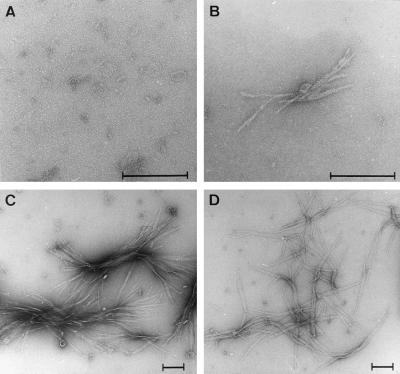
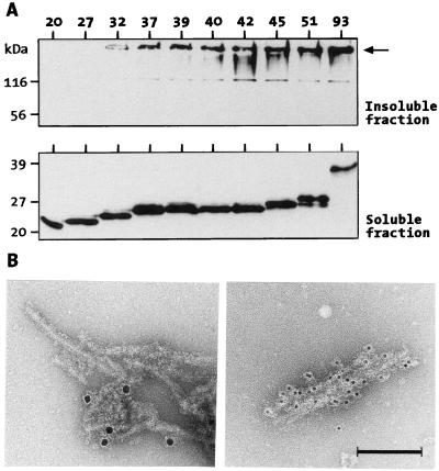
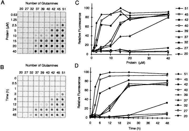
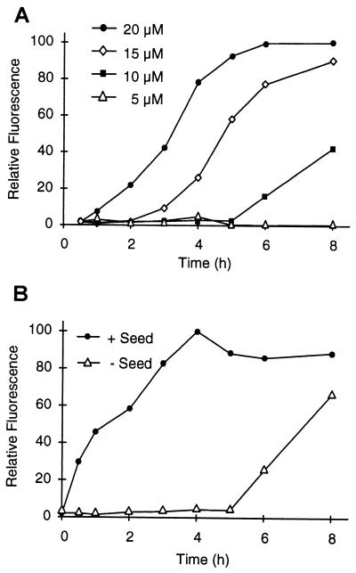
Huntington's disease is a progressive neurodegenerative disorder caused by a polyglutamine [poly(Q)] repeat expansion in the first exon of the huntingtin protein. Previously, we showed that N-terminal huntingtin peptides with poly(Q) tracts in the pathological range (51-122 glutamines), but not with poly(Q) tracts in the normal range (20 and 30 glutamines), form high molecular weight protein aggregates with a fibrillar or ribbon-like morphology, reminiscent of scrapie prion rods and beta-amyloid fibrils in Alzheimer's disease. Here we report that the formation of amyloid-like huntingtin aggregates in vitro not only depends on poly(Q) repeat length but also critically depends on protein concentration and time. Furthermore, the in vitro aggregation of huntingtin can be seeded by preformed fibrils. Together, these results suggest that amyloid fibrillogenesis in Huntington's disease, like in Alzheimer's disease, is a nucleation-dependent polymerization.


Figures

Figure 1
Figure 1
SDS/PAGE analysis of purified GST-HDex1 proteins and their tryptic digestion products. (A) Structure of the GST fusion proteins showing potential factor Xa (X) and trypsin (T) cleavage sites. The amino acid sequence corresponding to exon 1 of huntingtin is shown in bold. (B) SDS/12.5% PAGE gel stained with Coomassie blue. Five micrograms of each fusion protein, containing poly(Q) tracts of 20, 27, 32, 39, 40, 42, 45, or 51 residues, was loaded in each lane. (C and D) Immunoblot analysis of GST-HDex1 proteins before (C) and after (D) trypsin digestion. Incubation with trypsin was carried out for 24 h at a protein concentration of 0.8 mg/ml. Eighty nanograms of each fusion protein was loaded in each lane. ← marks the origin of electrophoresis.
Figure 2
Figure 2
Electron microscope pictures of HDex1 fibrils. Samples of the trypsin-digested GST-HDex1 proteins analyzed in Fig. 1D were negatively stained with 1% uranyl acetate and viewed by using electron microscopy. (A) HDex1-Q27. (B) HDex1-Q32. (C) HDex1-Q37. (D) HDex1-Q45. (Bar = 200 nm.)
Figure 3
Figure 3
(A) HDex1p expression and aggregation in COS cells. COS-1 cells transfected with HDex1-Q20, -Q27, -Q32, -Q37, -Q39, -Q40, -Q45, -Q51, and -Q93 constructs were harvested 42 h after transfection and lysed, and the crude cell lysates were separated by centrifugation into an insoluble (pellet) and a soluble (supernatant) fraction (see Materials and Methods). Ten micrograms of total protein from each soluble fraction and 10 μl of each insoluble fraction were run on separate SDS gels and immunoblotted with anti-HD1 serum. ← in the Upper gel marks the origin of electrophresis. (B) Electron micrographs of negatively stained immunogold-labeled HDex1p aggregates. (Left) Protein aggregates formed from purified GST-HDex1-Q51 protein after cleavage with factor Xa. (Right) Aggregated HDex1 protein isolated from HDex1-Q51-expressing COS-1 cells. The sizes of the gold particles were 10 and 5 nm, respectively. (Bar = 100 nm.)
Figure 4
Figure 4
(A) Concentration dependence of HDex1p aggregation. GST-HDex1 proteins with poly(Q) tracts of different lengths were incubated at the indicated concentrations with trypsin for 24 h at 37°C. Aliquots (200 ng) of each protein were then diluted into 0.2 ml of 2% SDS/50 mM DTT, boiled for 3 min, and filtered through a cellulose acetate membrane. Captured aggregates were detected by incubation with anti-AG51 serum (1:1,000), followed by incubation with alkaline phosphatase-conjugated anti-rabbit secondary antibody and the fluorescent substrate AttoPhos. (B) Time course of HDex1p aggregation. The various GST-HDex1 proteins were incubated at a concentration of 20 μM with trypsin. At the indicated times, aliquots (200 ng) of each protein were removed and analyzed by the filter retardation assay as in A. (C and D) Quantitative analysis of the dot-blot results shown in A and B, respectively. The relative amount of aggregate for each sample was quantitated on a Fuji-Imager (LAS 2000). For each experiment, the dot with the highest signal intensity was arbitrarily set as 100. The data reported are representative for three independent experiments using, in part, different GST-HDex1p preparations.
Figure 5
Figure 5
HDex1p aggregation requires a nucleation event. The aggregation assay was performed as in Fig. 4 by using dot-blot filtration on a cellulose acetate membrane and subsequent immunodetection of the captured aggregates. For each experiment shown, 100% represents maximum fluorescence intensity. (A) Effect of protein concentration on the kinetics of HDex1p aggregation. GST-HDex1-Q45 at 5, 10, 15, and 20 μM was incubated with trypsin at 37°C. At the indicated times, aliquots (200 ng) of each protein mixture were removed, denatured and reduced, and filtered. (B) HDex1p aggregate formation can be seeded by preformed fibrils. HDex1p aggregation was performed with GST-HDex1-Q45 at 10 μM in the presence or absence of added HDex1-Q45 fibrils (0.15 equivalent, 1.5 μM). A sample containing the seed alone was incubated in parallel, and the amount of aggregate captured by filtration at each time point was subtracted from the reported values. The results shown are from a single experiment performed in duplicate.

References

    1. Becher M W, Kotzuk J A, Sharp A H, Davies S W, Bates G P, Price D L, Ross C A. Neurobiol Dis. 1997;4:1–11. - PubMed
    1. DiFiglia M, Sapp E, Chase K O, Davies S W, Bates G P, Vonsattel J P, Aronin N. Science. 1997;277:1990–1993. - PubMed
    1. Igarashi S, Koide R, Shimohata T, Yamada M, Hayashi Y, Takano H, Date H, Oyake M, Sato T, Sato A, et al. Nat Genet. 1998;18:111–117. - PubMed
    1. Li M M, Kobayashi Y, Merry D E, Yamamoto M, Tanaka F, Doyu M, Hashizume Y, Fischbeck K H, Sobue G. Ann Neurol. 1998;44:249–254. - PubMed
    1. Skinner P J, Koshy B T, Cummings C L, Klement I A, Helin K, Servadio A, Zoghbi H Y, Orr H T. Nature (London) 1997;389:971–974. - PubMed

MeSH terms