. 1999 Apr 13;96(8):4639-44.
doi: 10.1073/pnas.96.8.4639.

Expression of the antiproliferative gene TIS21 at the onset of neurogenesis identifies single neuroepithelial cells that switch from proliferative to neuron-generating division

Affiliations

Expression of the antiproliferative gene TIS21 at the onset of neurogenesis identifies single neuroepithelial cells that switch from proliferative to neuron-generating division

P Iacopetti et al. Proc Natl Acad Sci U S A. .

Abstract

At the onset of mammalian neurogenesis, neuroepithelial (NE) cells switch from proliferative to neuron-generating divisions. Understanding the molecular basis of this switch requires the ability to distinguish between these two types of division. Here we show that in the mouse ventricular zone, expression of the mRNA of the antiproliferative gene TIS21 (PC3, BTG2) (i) starts at the onset of neurogenesis, (ii) is confined to a subpopulation of NE cells that increases in correlation with the progression of neurogenesis, and (iii) is not detected in newborn neurons. Expression of the TIS21 mRNA in the NE cells occurs transiently during the cell cycle, i.e., in the G1 phase. In contrast to the TIS21 mRNA, the TIS21 protein persists through the division of NE cells and is inherited by the neurons, where it remains detectable during neuronal migration and the initial phase of differentiation. Our observations indicate that the TIS21 gene is specifically expressed in those NE cells that, at their next division, will generate postmitotic neurons, but not in proliferating NE cells. Using TIS21 as a marker, we find that the switch from proliferative to neuron-generating divisions is initiated in single NE cells rather than in synchronized neighboring cells.


Figures

Figure 1
Figure 1
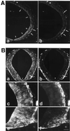
Characterization of the TIS21-antibodies. (A) Sequence comparison of the C-terminal one-third of mouse TIS21 (8) and mouse BTG1 (17). Amino acid residues identical between the two proteins are shaded. The sequences corresponding to the synthetic peptides used for immunization are boxed. The arrow indicates the single lysine residue in the K4 peptide that would be modified by glutaraldehyde-mediated coupling to keyhole limpet hemocyanin, thereby increasing the probability of the antibodies being directed against the C-terminal portion of the K4 peptide. The arrowhead indicates the alanine residue that is substituted by a threonine residue in rat TIS21 and mouse BTG1. (B) Western blot analysis of a cleared lysate of COS7 cells transfected with the TIS21 expression vector (+) or mock-transfected (−), using affinity-purified K4 antibody (1 μg/ml) or 1511 antiserum (1:2000). Arrowheads, TIS21; the dot indicates a TIS21-related 26 kDa protein detected upon overexpression of TIS21 in COS7 cells. (C) Immunoprecipitation from a total protein fraction of E12 mouse embryonic brain metabolically labeled for 1.5 h with [35S]methionine/cysteine, using affinity-purified K4 (5 μg/ml) or 1511 (9 μg/ml) antibody in the absence or presence of 50 μM K4 or 10 μM 1511 peptide as indicated. Note that in the two left lanes the background bands >20 kDa are also seen in the absence of antibody. To lower this background, the 35S-labeled total protein fraction used for immunoprecipitation in the six lanes on the right had been cleared by preadsorption to protein A-Sepharose before incubation with antibodies, and the immune complexes were eluted from protein A-Sepharose using 20 mM HCl instead of sample buffer. Arrowheads, TIS21. (D) Immunofluorescence of four adjacent (Da–Dd) 6-μm transverse cryosections through the hindbrain of an E11 mouse embryo, using the following affinity-purified primary antibodies: (Da) K4 antibody (0.5 μg/ml), (Db) K4 antibody preincubated with K4 peptide, (Dc) 1511 antibody (1.9 μg/ml), (Dd) 1511 antibody preincubated with 1511 peptide. The apical (ventricular) side of the neuroepithelium is up. Da vs. Db, and Dc vs. Dd, are the same length of exposure, and DaDd are the same magnification. [Bar = 85 μm (Dd).]
Figure 2
Figure 2
Comparison of the cellular distribution of the TIS21 mRNA (purple, arrowheads) and the neuronal marker βIII-tubulin (brown, arrows) in the E10 mouse brain by combined ISH/immunoperoxidase staining on cryosections. (A) Transverse section of the diencephalon at the level of the optic stalk. (B) Transverse section of the hindbrain. (C) Higher magnification of a portion of the field shown in A. (D) Higher magnification of a portion of the field shown in B. [Bar = 62 μm (A and B) or 20 μm (C and D).]
Figure 3
Figure 3
Correlation between the abundance of TIS21 mRNA-containing NE cells and newborn neurons in various regions of the E10 mouse brain. Sections subjected to combined ISH for TIS21 mRNA/immunoperoxidase staining for βIII-tubulin as in Fig. 2 were analyzed for the number of TIS21 mRNA-containing cells in the neuroepithelium and for the number of βIII-tubulin-positive cells in the mantle zone basal to the area of neuroepithelium analyzed. For βIII-tubulin-positive cells, only cells whose cell body showed immunoreactivity were considered. The number of βIII-tubulin-positive cells in the mantle zone is therefore underestimated, as is the number of TIS21 mRNA-containing cells in the neuroepithelium given the transient expression of the TIS21 mRNA in the G1 phase of the cell cycle (see Fig. 8 below). For each brain region, the numbers shown refer to a total area (neuroepithelium plus mantle zone) of 40,000 μm2 and were obtained by analysis of independent sections of the diencephalon (four sections), midbrain (two sections), and hindbrain (two sections).
Figure 4
Figure 4
Comparison of the cellular distribution of the TIS21 protein (Aa, Ba, Bc, and Be) and βIII-tubulin (Ab, Bb, Bd, and Bf) in the E10 mouse CNS by double immunofluorescence on transverse polyester sections through the midbrain (A) and spinal cord at the thoracical level (B). (A) Ventral and dorsal region, lower and upper portion of micrographs, respectively; arrows and arrow with asterisk, respectively, indicate neurons at the basal side of the neuroepithelium (Aa and Ab); arrowheads and arrowheads with asterisks, respectively, indicate TIS21 immunoreactivity in the neuroepithelium, which is concentrated toward the apical surface (Aa). White diamonds at the top of Aa and Ab, autofluorescent red blood cells. (B) White triangles at the bottom of Ba and Bb, floor plate. (Bc–Bf) the apical side of the neuroepithelium is on the left and the mantle zone is on the right. (Be and Bf) The ventral side; (Bc and Bd) more dorsal. Bar (in Bf) = 85 μm (Aa, Ab, Ba, and Bb) or 27 μm (Bc–Bf).
Figure 5
Figure 5
Comparison of the cellular distribution of the TIS21 mRNA (purple) and TIS21 protein (brown) in the E10 mouse brain by combined ISH/immunoperoxidase staining on cryosections. (A) Transverse section of the diencephalon at the level of the optic stalk. (B) Transverse section of the hindbrain. (C) Higher magnification of a portion of the field shown in A. (D) Higher magnification of a portion of the field shown in B. TIS21 mRNA expression is confined to the perikaryon of cells in the neuroepithelium (arrowheads); these cells also contain TIS21 protein in the perikaryon and, more prominently, in cell processes (for a comparison with cells stained for the TIS21 mRNA but not protein, see Fig. 2). Cells containing TIS21 protein but not mRNA are also observed in the neuroepithelium (open arrows). TIS21 protein but not mRNA is detected in neurons in the mantle zone (arrows). [Bar (in D) = 62 μm (A and B) or 20 μm (C and D).]
Figure 6
Figure 6
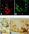
Analysis of newborn, migrating neurons in the ventricular zone of the E10 mouse brain for the expression of TIS21 mRNA and protein. (A and B) Double immunofluorescence for TIS21 (A) and βIII-tubulin (B) of a transverse polyester section through the spinal cord. Arrows, newborn neurons; dotted white lines, neuronal process emerging from perikaryon; arrowheads, NE cells showing TIS21 immunoreactivity concentrated toward the apical surface (A) but lacking βIII-tubulin (B). Asterisk in B, floor plate. (C and D) Combined ISH for TIS21 mRNA (purple)/immunoperoxidase staining for βIII-tubulin (brown) of transverse cryosections through the hindbrain. The apical side of the neuroepithelium is up. Arrows, newborn neurons in the ventricular zone; arrow with asterisk in D, newborn neuron in close proximity to the apical surface; arrowheads, NE cells that express TIS21 mRNA but lack βIII-tubulin immunoreactivity (for a comparison with cells showing both, an ISH and an immunoperoxidase signal, see Fig. 5 C and D, arrowheads). [Bar (in D) = 31 μm (A and B) or 20 μm (C and D).]
Figure 7
Figure 7
Comparison of the expression of the TIS21 mRNA and protein to markers of the cell cycle in the ventricular zone of the E10 mouse brain. (A and B) Double immunofluorescence for TIS21 (A) and the mitotic marker MPM-2 (B) of a transverse polyester section through the hindbrain. Some mitotic cells stained by the MPM-2 antibody also contain TIS21 (arrowheads), whereas other mitotic cells do not (arrows). (C–E) Double immunofluorescence confocal microscopy for TIS21 (C, red) and MPM-2 (E, green) of a transverse cryosection through the hindbrain. A single optical section is shown. (D) overlay. (F and G) Double immunofluorescence confocal microscopy for TIS21 (red) and BrdUrd (green) of transverse cryosections through the hindbrain of embryos labeled with BrdUrd for either 30 min (F) or 2 h (G). Single optical sections are shown. BrdUrd is localized to the nucleus, whereas the TIS21 antibody stains the cytoplasm. Examples of double-positive cells are outlined in white. Because the TIS21 protein is localized to the apical processes and ends of NE cells, BrdUrd-labeled nuclei could only be assigned to TIS21-positive cells when the former were sufficiently close to the apical surface, which is the case in late S phase and G2 phase because of interkinetic nuclear migration (22). For this reason, we could not quantitate the proportion of TIS21 protein-positive NE cells that contain S phase and G2 phase BrdUrd-labeled nuclei. (H1 and H2) Both panels show combined ISH for TIS21 mRNA (purple)/ immunoperoxidase staining for MPM-2 (brown) of transverse cryosections through the hindbrain. (I and J) Combined ISH for TIS21 mRNA (purple)/immunoperoxidase staining for BrdUrd (brown) of transverse cryosections through the hindbrain (I) or the midbrain (J) of embryos labeled with BrdUrd for either 30 min (I) or 2 h (J). The insets show double-positive cells (arrowheads) that are occasionally observed; in these cells, the brown outline of the nucleus is visible through the purple staining of the cell body. (A–J) The apical side of the neuroepithelium is up. [Bar (in J) = 17 μm (A and B), 13 μm (C–E), 25 μm (G), and 20 μm (F and H–J).]
Figure 8
Figure 8
Expression of the TIS21 gene in neuron-generating NE cells. See Discussion.
Figure 9
Figure 9
Analysis of TIS21 mRNA expression at the onset of neurogenesis by in situ hybridization on three serial (A–C), 5-μm transverse cryosections through the diencephalon of an E10 embryo. The example shown is representative of six independent observations. (Bar = 20 μm.)

Similar articles

Cited by

References

    1. Rakic P. Science. 1988;241:170–176. - PubMed
    1. McConnell S K. Neuron. 1995;15:761–768. - PubMed
    1. Takahashi T, Nowakowski R S, Caviness V S., Jr J Neurosci. 1996;16:6183–6196. - PMC - PubMed
    1. Huttner W B, Brand M. Curr Opin Neurobiol. 1997;7:29–39. - PubMed
    1. Cameron H A, Hazel T G, McKay R D G. J Neurobiol. 1998;36:287–306. - PubMed

Publication types

MeSH terms

LinkOut - more resources