. 1999 Apr 13;96(8):4656-61.
doi: 10.1073/pnas.96.8.4656.

Genetic background changes the pattern of forebrain commissure defects in transgenic mice underexpressing the beta-amyloid-precursor protein

Affiliations

Genetic background changes the pattern of forebrain commissure defects in transgenic mice underexpressing the beta-amyloid-precursor protein

F Magara et al. Proc Natl Acad Sci U S A. .

Abstract

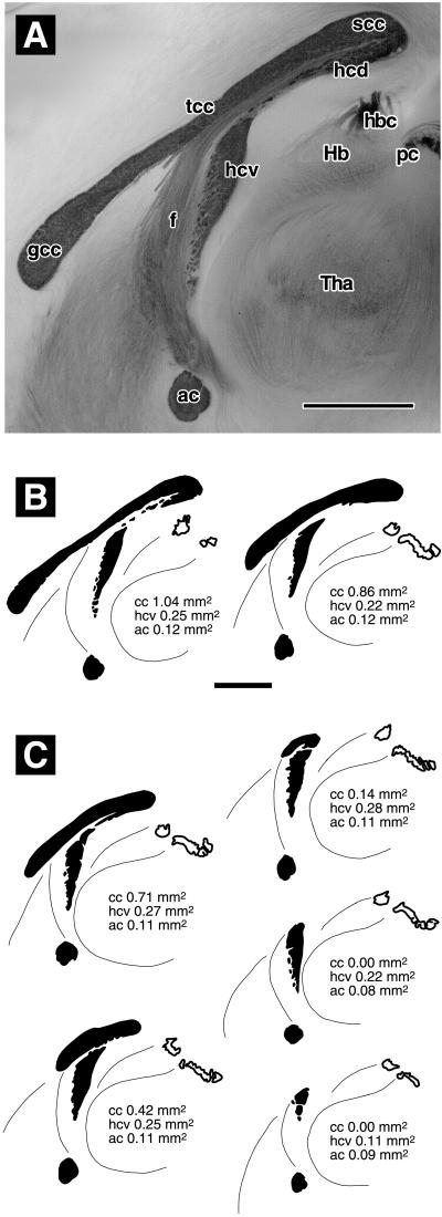
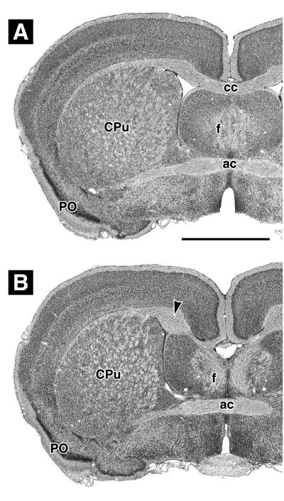
We previously have reported corpus callosum defects in transgenic mice expressing the beta-amyloid precursor protein (betaAPP) with a deletion of exon 2 and at only 5% of normal levels. This finding indicates a possible involvement of betaAPP in the regulation or guidance of axon growth during neural development. To determine to what degree the betaAPP mutation interacts with genetic background alleles that predispose for forebrain commissure defects in some mouse lines, we have assessed the size of the forebrain commissures in a sample of 298 mice. Lines with mixed genetic background were compared with congenic lines obtained by backcrossing to the parental strains C57BL/6 and 129/SvEv. Mice bearing a null mutation of the betaAPP gene also were included in the analysis. We show that, independently of genetic background, both lack and underexpression of betaAPP are associated with reduced brain weight and reduced size of forebrain commissures, especially of the ventral hippocampal commissure. In addition, both mutations drastically increase the frequency and severity of callosal agenesis and hippocampal commissure defects in mouse lines with 129/SvEv or 129/Ola background.


Figures

Figure 1
Figure 1
Classification of forebrain commissures in brains that were split in the midsagittal plane and stained with gold chloride. (A) C57BL/6 mouse with normal commissures. The anterior (ac) and ventral hippocampal (hcv) commissures are darkly stained, as are the genu (gcc), truncus (tcc), and splenium (scc) of the corpus callosum. There is no clear boundary between the dorsal hippocampal commissure (hcd) and the splenium. Fibers of the fornix (f) stain more weakly. They emerge along the ventral surface of the truncus, then run ventrally and rostrally parallel to the sectioning plane. Hb, Habenula; hbc, habenular commissure; pc, posterior commissure; Tha, thalamus. (B) Representative camera lucida drawings of commissures classified as normal: (Left) C57BL/6 (same brain as in A), (Right) a wild type of line βAPPΔ/Δ Sv. (C) Representative camera lucida drawings of commissures classified as abnormal. From top left to bottom right: slightly reduced corpus callosum (cc), strongly reduced corpus callosum, rudimentary corpus callosum, absent corpus callosum with still normal ventral hippocampal commissure, and absent corpus callosum with reduced ventral hippocampal commissure. (Bar in A = 1 mm. Bar in B = 1 mm, applies to B and C.) See text for details.
Figure 2
Figure 2
Cresyl violet-stained coronal sections through plastic-embedded brains, taken at a level showing the anterior commissure (ac), fornix columns (f), caudatoputamen (CPu), and primary olfactory cortex (PO). (A) βAPPΔ/Δ B6-Sv mouse with normal corpus callosum. (B) βAPPΔ/Δ Sv mouse with missing corpus callosum and well-developed Probst bundles (arrowhead). (Bar in A = 1 mm, applies to A and B.) See text for details.

Similar articles

Cited by

References

    1. Livy D J, Wahlsten D. J Hered. 1991;82:459–464. - PubMed
    1. Wahlsten D. Brain Res. 1974;68:1–18. - PubMed
    1. Probst M. Arch Psychiatr Nervenkr. 1901;34:709–786.
    1. Ozaki H S, Shimada M. Brain Res. 1988;441:5–14. - PubMed
    1. Livy D J, Wahlsten D. Hippocampus. 1997;7:2–14. - PubMed

Publication types

Substances