. 1999 Apr 13;96(8):4692-7.
doi: 10.1073/pnas.96.8.4692.

Stable expression of human beta1,4-galactosyltransferase in plant cells modifies N-linked glycosylation patterns

Affiliations

Stable expression of human beta1,4-galactosyltransferase in plant cells modifies N-linked glycosylation patterns

N Q Palacpac et al. Proc Natl Acad Sci U S A. .

Abstract

beta1,4-Galactosyltransferase (UDP galactose: beta-N-acetylglucosaminide: beta1,4-galactosyltransferase; EC 2.4.1. 22) catalyzes the transfer of galactose from UDP-Gal to N-acetylglucosamine in the penultimate stages of the terminal glycosylation of N-linked complex oligosaccharides in mammalian cells. Tobacco BY2 cells lack this Golgi enzyme. To determine to what extent the production of a mammalian glycosyltransferase can alter the glycosylation pathway of plant cells, tobacco BY2 suspension-cultured cells were stably transformed with the full-length human galactosyltransferase gene placed under the control of the cauliflower mosaic virus 35S promoter. The expression was confirmed by assaying enzymatic activity as well as by Southern and Western blotting. The transformant with the highest level of enzymatic activity has glycans with galactose residues at the terminal nonreducing ends, indicating the successful modification of the plant cell N-glycosylation pathway. Analysis of the oligosaccharide structures shows that the galactosylated N-glycans account for 47.3% of the total sugar chains. In addition, the absence of the dominant xylosidated- and fucosylated-type sugar chains confirms that the transformed cells can be used to produce glycoproteins without the highly immunogenic glycans typically found in plants. These results demonstrate the synthesis in plants of N-linked glycans with modified and defined sugar chain structures similar to mammalian glycoproteins.


Figures
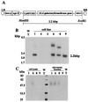
Figure 1
Figure 1
Expression of human β1,4-galactosyltransferase (hGT) gene in tobacco BY2 cells. (A) The recombinant hGT plasmid for expression of human β1,4-galactosyltransferase in tobacco cells. The hGT coding region lies downstream of the CaMV35S promoter, followed by the nopaline synthase terminator (nos-t) from pBI221. The construct also has the neomycin phosphotransferase II (nptII) gene under the control of the nopaline synthase promoter (nos-p) from pGA482. RB/LB, right and left borders, respectively. (B) Southern blot analyses of genomic DNAs from transformed and wild-type BY2 tobacco cells. DNAs were digested with HindIII and EcoRI and probed with a 32P-labeled hGT gene fragment as described in Materials and Methods. WT, wild type; 1, 4, 5, 6, 8, and 9, cell line number for transformed tobacco cells. The numbers to the left indicate the sizes (kbp) and positions of λ DNA fragments digested with HindIII. Size of the hybridizing fragment (2.2 kbp) is indicated. (C) Western blots of immunoreactive proteins from transgenic and nontransgenic tobacco cells. Proteins were denatured, resolved by SDS/PAGE, and electroblotted onto a nitrocellulose membrane. The blots were probed with anti-hGT antibody as described in Materials and Methods. Lanes contain proteins from cell extracts of cell lines 1, 6, 8, 9 and wild type and microsome fractions of cell lines 1, 6, 8, 9 and wild type. Molecular mass of marker proteins are indicated on the left.
Figure 2
Figure 2
Detection of galactosylated glycoproteins by using R. communis RCA120 affinity chromatography. Eluted fractions were subjected to SDS/PAGE, and the gel was visualized by silver staining (A) or blotted onto a nitrocellulose membrane and lectin (RCA120) stained (B). Lanes 1 and 2, proteins from wild-type BY2 (WT); 3 and 4, proteins from transformed GT6. (C) Blots probed with a xylose-specific antiserum for plant complex glycans. Lanes 1 and 2, total protein extracts from BY2 and GT6; 3, glycoproteins from GT6 after RCA120 affinity chromatography. Molecular mass markers are in kDa.
Figure 3
Figure 3
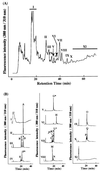
PA derivatives from glycoproteins expressed in transformed cells. (A) RP-HPLC pattern of PA-sugar chains eluted by increasing the acetonitrile concentration in 0.02% trifluoroacetic acid linearly from 0 to 15% for 60 min at a flow rate of 1.2 ml/min. I–XI, individual fractions collected and purified in SF-HPLC. (B) SF-HPLC of PA-sugar chains in A. PA-sugar chains were eluted by increasing the water content in the water-acetonitrile mixture from 30 to 50% for 40 min at a flow rate of 0.8 ml/min. Excitation and emission wavelengths were 310 and 380 nm, respectively. Fractions that contain N-linked sugar chains are marked with ∗.
Figure 4
Figure 4
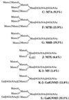
Proposed structures of N-linked glycans obtained from transformed cells. Enclosed numbers in parentheses represent molar ratios.
Figure 5
Figure 5
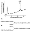
Elution position of peak K-2 in RP-HPLC compared with two authentic sugar chains, A and B. Elution conditions are described as in Fig. 3A.
Figure 6
Figure 6
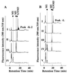
SF-HPLC profiles of galactosylated PA-sugar chains obtained after exoglycosidase digestions. PA-sugar chains were eluted by increasing the water content in the water-acetonitrile mixture from 30 to 50% for 25 min at a flow rate of 0.8 ml/min. (A) PA-sugar chain K-2. I, elution position of native galactosylated PA-sugar chain; II, β-galactosidase digest of I; III, β-N-acetylglucosaminidase digest of II; IV, jackbean α-mannosidase digest of III. (B) PA-sugar chain L. I, elution position of native galactosylated PA-sugar chain; II, β-galactosidase digest of I; III, β-N-acetylglucosaminidase digest of II; IV, α1,2 mannosidase digest of III; V, jackbean α-mannosidase digest of III.

Similar articles

Cited by

References

    1. Cramer C L, Weissenborn D L, Oishi K K, Grabau E A, Bennett S, Ponce E, Grabowski G A, Radin D N. Ann N Y Acad Sci. 1996;792:62–71. - PubMed
    1. Kusnadi A R, Nikolov Z L, Howard J A. Biotechnol Bioeng. 1997;56:473–484. - PubMed
    1. Arntzen C J. Nat Biotechnol. 1997;15:221–222. - PubMed
    1. Pen J. In: Transgenic Plants: A Production System for Industrial and Pharmaceutical Proteins. Owen M R L, Pen J, editors. New York: Wiley; 1996. pp. 149–167.
    1. Kukuruzinska M A, Lennon K. Crit Rev Oral Biol Med. 1998;9:415–448. - PubMed

LinkOut - more resources