. 1999 Apr 13;96(8):4718-23.
doi: 10.1073/pnas.96.8.4718.

Genes for calcineurin B-like proteins in Arabidopsis are differentially regulated by stress signals

Affiliations

Genes for calcineurin B-like proteins in Arabidopsis are differentially regulated by stress signals

J Kudla et al. Proc Natl Acad Sci U S A. .

Abstract

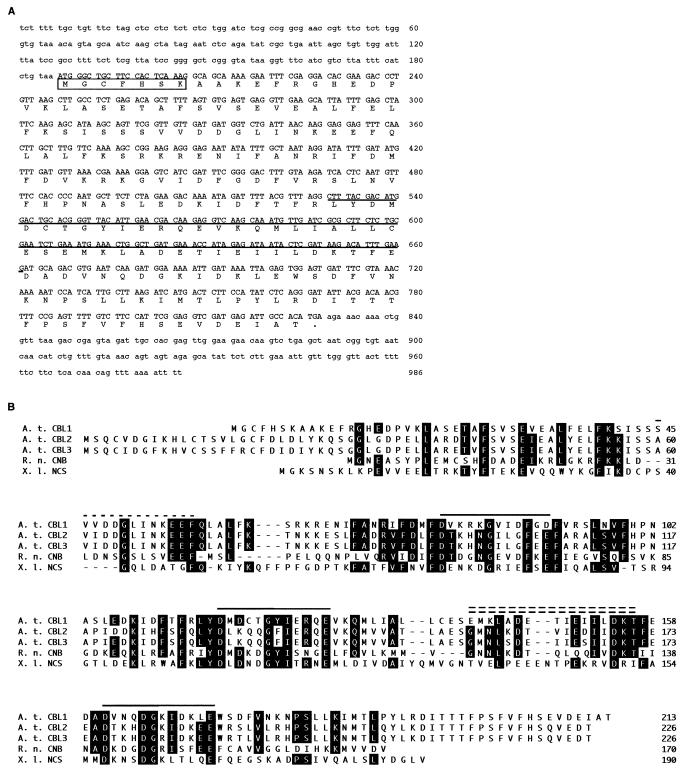
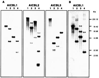
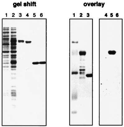
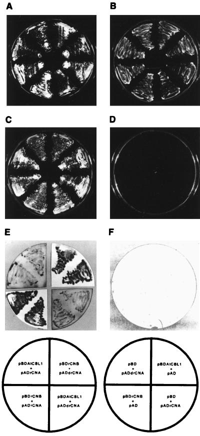
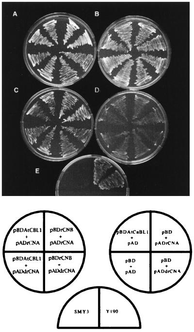
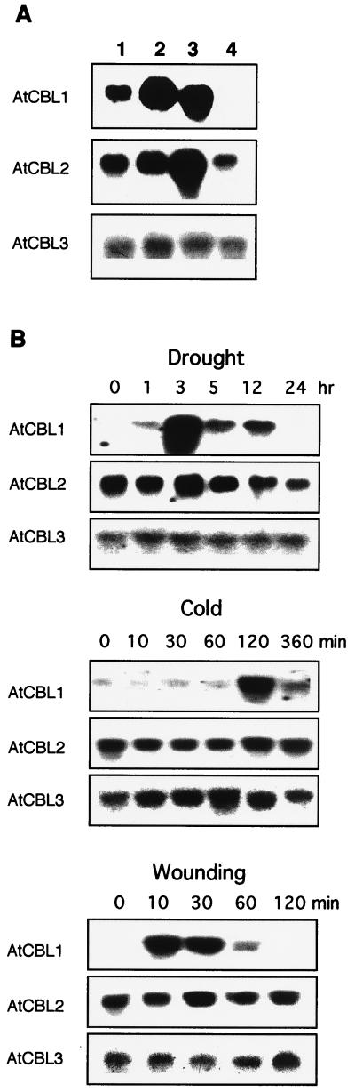
An important effector of Ca2+ signaling in animals and yeast is the Ca2+/calmodulin-dependent protein phosphatase calcineurin. However, the biochemical identity of plant calcineurin remained elusive. Here we report the molecular characterization of AtCBL (Arabidopsis thaliana calcineurin B-like protein) from Arabidopsis. The protein is most similar to mammalian calcineurin B, the regulatory subunit of the phosphatase. AtCBL also shows significant similarity with another Ca2+-binding protein, the neuronal calcium sensor in animals. It contains typical EF-hand motifs with Ca2+-binding capability, as confirmed by in vitro Ca2+-binding assays, and it interacts in vivo with rat calcineurin A in the yeast two-hybrid system. Interaction of AtCBL1 and rat calcineurin A complemented the salt-sensitive phenotype in a yeast calcineurin B mutant. Cloning of cDNAs revealed that AtCBL proteins are encoded by a family of at least six genes in Arabidopsis. Genes for three isoforms were identified in this study. AtCBL1 mRNA was preferentially expressed in stems and roots and its mRNA levels strongly increased in response to specific stress signals such as drought, cold, and wounding. In contrast, AtCBL2 and AtCBL3 are constitutively expressed under all conditions investigated. Our data suggest that AtCBL1 may act as a regulatory subunit of a plant calcineurin-like activity mediating calcium signaling under certain stress conditions.


Figures

Figure 1
Figure 1
Sequence analyses of AtCBL1 cDNA and AtCBL proteins. (A) cDNA and deduced amino acid sequences of AtCBL1. The coding region of the cDNA is presented in capital letters and the noncoding regions in lowercase letters. The boxed sequence contains a typical myristoylation site (MGXXXSK). The underlined sequence represents the cDNA fragment identified by PCR (see Materials and Methods). An in-frame stop codon (taa) is immediately upstream from the ATG starting codon. (B) Alignment of the amino acid sequences of three AtCBL proteins from Arabidopsis (A. t. CBL1, A. t. CBL2, A. t. CBL3) with rat CNB (R. n. CNB), and Xenopus neuronal calcium sensor (X. l. NCS). Alignment was generated by using the clustal method with dnastar software. Amino acids identical to the consensus sequence of the alignment are shown on black background. The numbers on the right indicate the amino acid position. Dashes indicate gaps introduced to improve the alignment. Solid lines above the sequence indicate position of EF-hand regions. The single-dashed line represents a variation of the Ca2+-binding domain. The double-dashed line denotes the CNA interaction domain.
Figure 2
Figure 2
Genomic organization of AtCBL genes. Aliquots (5 μg per digestion) of genomic DNA from Arabidopsis plants (Columbia ecotype) were digested with BamHI (lanes 1), BglII (lanes 2), EcoRI (lanes 3), and HindIII (lanes 4). (A) After agarose gel electrophoresis, DNA was blotted onto nylon membranes and hybridized under high-stringency conditions with 32P-labeled probes for AtCBL1, AtCBL2, and AtCBL3, respectively. (B) Low-stringency hybridization was performed using AtCBL1 cDNA as a probe. HindIII-digested phage λ DNA was used to provide molecular size markers.
Figure 3
Figure 3
AtCBL1 is a functional Ca2+-binding protein. In the gel-shift panel, lane 1 and lane 2 were loaded with protein extract prepared before and after isopropyl β-d-thiogalactopyranoside (IPTG) induction, respectively. GST-AtCBL1 fusion protein was purified and incubated in EGTA- (lane 3) or calcium-containing (lane 4) buffer before being analyzed by SDS/PAGE. As a negative control, GST protein was analyzed in the same way (lanes 5 and 6). In the overlay assay, lanes 1–3 show Coomassie blue-stained molecular weight marker, GST-AtCBL1 (0.4 μg), and GST (0.2 μg), respectively. Lanes 4–6 indicate the same samples transferred to the nitrocellulose membrane and assayed by 45Ca2+ binding. Only the fusion protein in lane 2 shows Ca2+-binding capability (lane 5).
Figure 4
Figure 4
Interaction between AtCBL1 and rat CNA protein in the yeast two-hybrid system. Growth of yeast transformants on SC −Trp −Leu medium (A and B), SC −Trp −Leu −His containing 25 mM 3-aminotriazole (C and D), and the colony color of the transformants determined by the filter lift assay (E and F) are shown. Plates containing histidine, but lacking tryptophan and leucine, select for the presence of the two plasmids containing the Gal4 DNA-binding and the Gal4 activation domain. Plates lacking tryptophan, leucine, and histidine and supplemented with 25 mM 3-aminotriazole select for a positive interaction between the fusion proteins. The array of the yeasts containing the different constructs is indicated in the scheme at the bottom. The symbols for the constructs were described in the text.
Figure 5
Figure 5
Complementation of yeast CNB mutant by interaction of rat CNA and AtCBL1. Yeast CNB mutant strain SMY3 (28) was transformed with various combinations of plasmids indicated in the circles. Annotations of the plasmids are shown in Materials and Methods and are the same as used in Fig. 4 and Table 1. The same combinations were used in A and C (left circle in the diagram at the bottom), and in B and D (right circle). Mutant (SMY3) and a wild-type (Y190) controls in E are shown in the half-circle. Yeast strains were grown on the synthetic medium containing 0 mM (A and B) and 200 mM (C, D, and E) LiCl.
Figure 6
Figure 6
Expression pattern of three members of AtCBL gene family. (A) mRNA levels in leaves (lane 1), stems (lane 2), roots (lane 3), and flowers (lane 4). (B) mRNA accumulation under the stress conditions drought, cold, and wounding. A 10-μg sample of total RNA was loaded into each lane for RNA gel analysis using AtCBL cDNAs as hybridization probes.

Comment in

  • How plants learn.
    Trewavas A. Trewavas A. Proc Natl Acad Sci U S A. 1999 Apr 13;96(8):4216-8. doi: 10.1073/pnas.96.8.4216. Proc Natl Acad Sci U S A. 1999. PMID: 10200239 Free PMC article. Review. No abstract available.

References

    1. Clapham D E. Cell. 1995;80:259–268. - PubMed
    1. Trewavas A, Read N, Campbell A K, Knight M. Biochem Soc Trans. 1996;24:971–974. - PubMed
    1. Hunter T. Cell. 1995;80:225–236. - PubMed
    1. Guerini D. Biochem Biophys Res Commun. 1997;235:271–275. - PubMed
    1. Klee C B, Draetta G F, Hubbard M J. Adv Enzymol. 1988;61:149–200. - PubMed

Publication types

MeSH terms

Associated data

LinkOut - more resources