. 1999 May 1;516 ( Pt 3)(Pt 3):875-83.
doi: 10.1111/j.1469-7793.1999.0875u.x.

Gating of transmission in climbing fibre paths to cerebellar cortical C1 and C3 zones in the rostral paramedian lobule during locomotion in the cat

Affiliations

Gating of transmission in climbing fibre paths to cerebellar cortical C1 and C3 zones in the rostral paramedian lobule during locomotion in the cat

R Apps et al. J Physiol. .

Abstract

1. Climbing fibre field potentials evoked by low intensity (non-noxious) electrical stimulation of the ipsilateral superficial radial nerve have been recorded in the rostral paramedian lobule (PML) in awake cats. Chronically implanted microwires were used to monitor the responses at eight different C1 and C3 zone sites during quiet rest and during steady walking on a moving belt. The latency and other characteristics of the responses identified them as mediated mainly via the dorsal funiculus-spino-olivocerebellar path (DF-SOCP). 2. At each site, mean size of response (measured as the area under the field, in mV ms) varied systematically during the step cycle without parallel fluctuations in size of the peripheral nerve volley. Largest responses occurred overwhelmingly during the stance phase of the step cycle in the ipsilateral forelimb while smallest responses occurred most frequently during swing. 3. Simultaneous recording from pairs of C1 zone sites located in the anterior lobe (lobule V) and C1 or C3 zone sites in rostral PML revealed markedly different patterns of step-related modulation. 4. The findings shed light on the extent to which the SOCPs projecting to different parts of a given zone can be regarded as functionally uniform and have implications as to their reliability as channels for conveying peripheral signals to the cerebellum during locomotion.


Figures

Figure 1
Figure 1. Cortical location of recording sites
Dorsal view of the unfolded cat cerebellar cortex (left hemicerebellum only) with expanded views to show the approximate location of individual microwire recording sites. A, 7 sites within the C1 zone in lobule Va-c of the anterior lobe and B, 8 sites within the C1 and C3 zones in the rostral paramedian lobule of the posterior lobe. AL, anterior lobe; PL, posterior lobe; PML, paramedian lobule; pf, primary fissure.
Figure 3
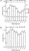
Figure 3. Step analysis for one rostral PML C1 zone site
In A and B the step cycle has been divided into tenths with bin one coinciding with the onset of locomotor EMG in the ipsilateral triceps brachii muscle. Periods of stance and swing are approximate timings for trajectory of the ipsilateral forelimb in this and subsequent figures. A, step histogram to show the mean size of the climbing fibre field potential in terms of its total area (+s.e.m.). Dashed horizontal line, mean size of response at rest. •-•, mean peak-to-peak amplitude of the nerve volley. B, same data but mean size of the response in terms of the area under the initial 2 ms of the field.
Figure 2
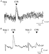
Figure 2. Example climbing fibre field potentials in rostral PML
A, extracellular recording from the granular layer of the cerebellar cortex (4 consecutive sweeps superimposed) to show a C1 zone climbing fibre field potential evoked by ipsilateral superficial radial nerve stimulation (intensity, 2T). B, same site to show the effect of paired stimuli (interstimulus interval of 30 ms, average of 4 sweeps). Stim, stimulus; CFR, climbing fibre response.
Figure 4
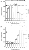
Figure 4. Simultaneous recording from a pair of rostral PML and lobule V sites
A and B, data obtained at 2 different recording sites evoked by the same ipsilateral superficial radial nerve stimulation (intensity, 2T). A, step histogram for a C1 zone site in rostral PML and B, step histogram for a C1 zone site in lobule V. Each histogram constructed according to the conventions of Fig. 3.
Figure 5
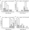
Figure 5. Frequency distributions for times of largest and smallest cerebellar responses
A shows distribution of the timing during the step cycle in the ipsilateral forelimb when mean size of response to SR stimulation was largest. Data obtained from 20 step histograms from 8 rostral PML recording sites. B, same as A, but for timing of smallest mean size of response. C, as A but for responses recorded in lobule V. Data obtained from 20 step histograms from 7 recording sites. D, as C but for timing of smallest mean size of response. Stippling selects for each recording site the timings from the step histogram with the least variation in nerve volley size.
Figure 6
Figure 6. Shifts in step timing between pairs of rostral PML and lobule V sites
A, pairs of rostral PML and lobule V step histograms generated by the same nerve stimuli were compared as to the extent of any shift in the position of the step tenth in which response mean size was largest (n = 13 comparisons). B, same as A but for shifts in step tenth for timing of the smallest response mean size.

Comment in

Similar articles

Cited by

References

    1. Apps R. Movement related gating of climbing fibre input to cerebellar cortical zones. Progress in Neurobiology. 1999;57:537–562. 10.1016/S0301-0082(98)00068-9. - DOI - PubMed
    1. Apps R, Atkins MJ, Garwicz M. Gating of cutaneous input to cerebellar climbing fibres during a reaching task in the cat. The Journal of Physiology. 1997;502:203–214. - PMC - PubMed
    1. Apps R, Hartell NA, Armstrong DM. Step phase-related excitability changes in spino-olivocerebellar paths to the c1 and c3 zones in cat cerebellum. The Journal of Physiology. 1995;483:687–702. - PMC - PubMed
    1. Apps R, Lee S. Movement-related gating of spino-olivocerebellar paths in the awake cat. The Journal of Physiology. 1997;504.P:16–17S.
    1. Apps R, Lidierth M, Armstrong DM. Locomotion-related variations in excitability of spino-olivocerebellar paths to cat cerebellar cortical c2 zone. The Journal of Physiology. 1990;424:487–512. - PMC - PubMed

Publication types

LinkOut - more resources