. 1999 May 1;516 ( Pt 3)(Pt 3):897-906.
doi: 10.1111/j.1469-7793.1999.0897u.x.

Modality-specific hyper-responsivity of regenerated cat cutaneous nociceptors

Affiliations

Modality-specific hyper-responsivity of regenerated cat cutaneous nociceptors

D Andrew et al. J Physiol. .

Abstract

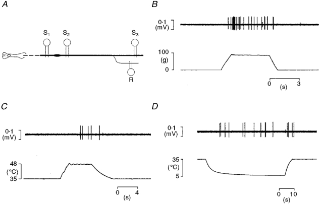
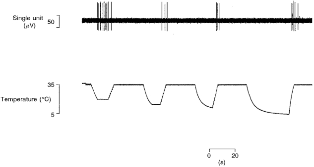
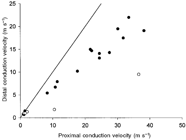
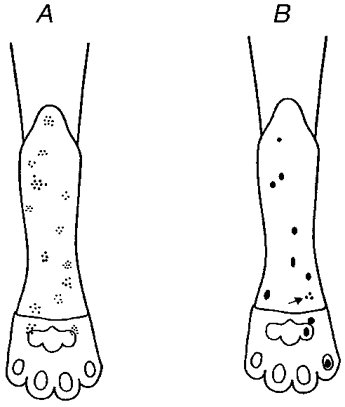
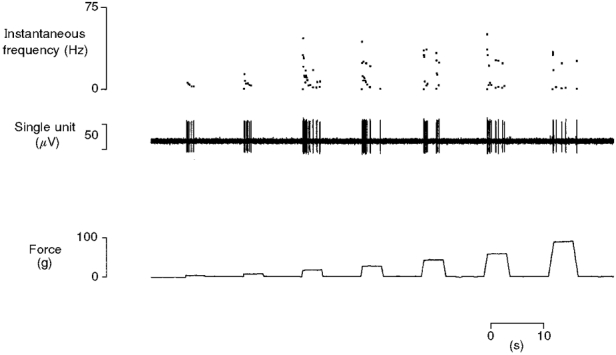
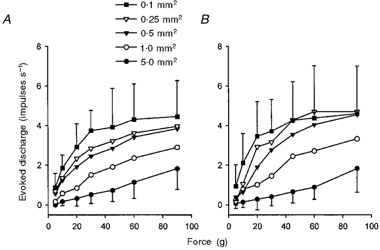
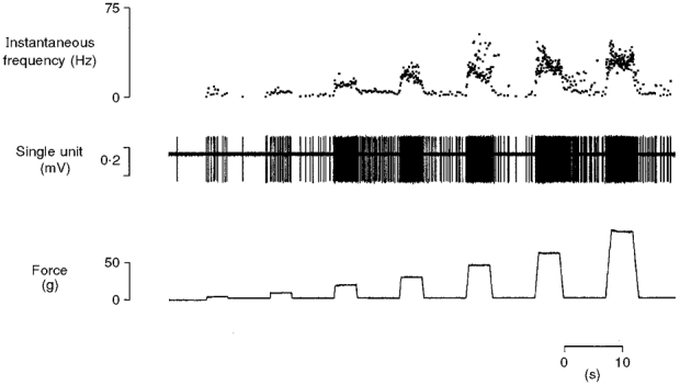
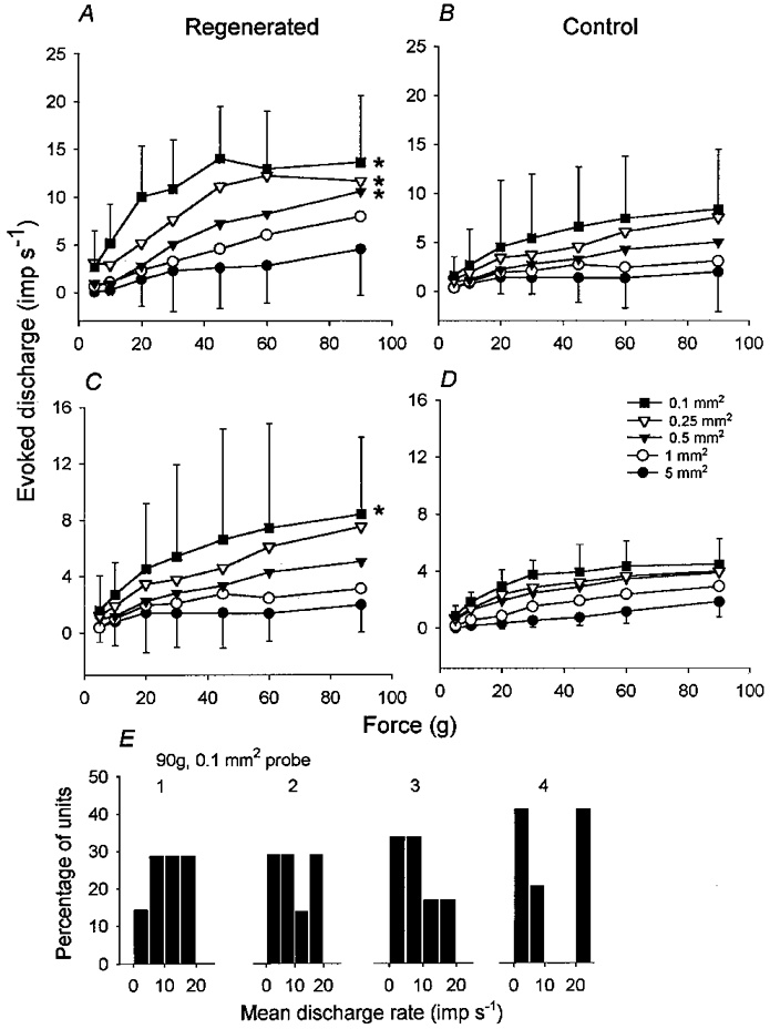
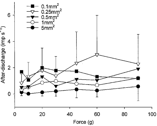
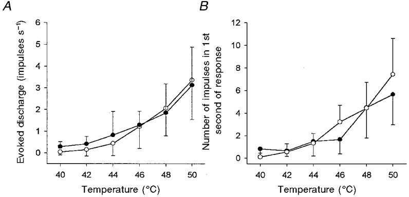
1. Experiments were performed on anaesthetized cats to investigate the receptive properties of regenerated cutaneous tibial nerve nociceptors, and to obtain evidence for coupling between them and other afferent fibres as being possible peripheral mechanisms involved in neuropathic pain. These properties were studied 6-7 months after nerve section and repair. 2. Recordings were made from 25 regenerated nociceptors; 14 were A fibres and the remainder were C fibres. Their receptive field sizes and conduction velocities were similar to controls. There was no significant difference between their mechanical thresholds and those of a control population of nociceptors. 3. Regenerated nociceptors were significantly more responsive to suprathreshold mechanical stimuli than were uninjured control fibres. This increase in mechanical sensitivity occurred in both A and C fibres, although A fibres showed a greater increase in mechano-sensitivity than C fibres. Over half of the regenerated nociceptors (13/25) showed after-discharge to mechanical stimuli which was never seen in controls; the mean firing rate during this period of after-discharge was significantly related to both stimulus intensity and stimulus area. 4. There was no significant difference between the heat encoding properties of regenerated nociceptors and control nociceptors. Cold sensitivity was similarly unchanged. Thus, abnormal peripheral sprouting was unlikely to account for the increased mechanical sensitivity of the regenerated fibres. None of the regenerated nociceptors were found to be coupled to other fibres. 5. These results suggest that the clinical observation of mechanical hyperalgesia in patients after nerve injury may have a peripheral basis. Based on this model, other signs of neuropathic pain (i.e. tactile or thermal allodynia) are more likely to be due to altered central processing.


Figures

Figure 1
Figure 1. Diagram of the preparation and testing the receptive properties of regenerated nociceptors
A, schematic representation of the preparation showing the sites of electrical stimulation and of recording of single units (S1-S3). B-D, examples of responses of a regenerated nociceptor (a mechano-heat-cold fibre, CV from S1 0.7 m s−1) to mechanical (B) and thermal (C and D) stimulation of its receptive field.
Figure 10
Figure 10. Cold sensitivity of control C fibre nociceptors
A typical example of cold-evoked responses from a saphenous nerve C fibre (CV 1.1 m s−1). The unit responded directly to a 20 °C stimulus from an adapting skin temperature of 35 °C but not to colder stimuli, it also responded reproducibly during rewarming.
Figure 2
Figure 2. Slowing of conduction velocity of regenerated myelinated nociceptors
Myelinated, but not unmyelinated regenerated nociceptors conduct faster proximal to the injury site than through it. Proximal conduction velocities were obtained from latency measurements evoked by stimulation at S2 (Fig. 1A). Distal conduction velocities were obtained from stimulation at S1. For convenience faster conduction velocities have been plotted on the horizontal axis. ○, units whose distal conduction velocities were only determined by electrocutaneous stimulation (see Methods). The continuous line is the line of unity.
Figure 3
Figure 3. Distribution of receptive fields of regenerated nociceptors
Receptive field distribution for regenerated A fibres (A) and regenerated C fibres (B). The arrow indicates a C fibre with a multi-spot receptor.
Figure 4
Figure 4. Typical response to mechanical stimuli of a control C fibre nociceptor
Mechanically evoked responses from a single C fibre (CV 1.1 m s−1) to progressively more intense stimuli delivered with a probe of tip area 0.25 mm2. The response of the unit increases with stimulus intensity. It is quiescent between stimuli and also shows the bursting typical of C fibres.
Figure 5
Figure 5. Intensity coding of mechanical stimuli by C fibre nociceptors in two different cutaneous fields
A, responses of 9 C fibres in the saphenous nerve to mechanical stimulation with probes of tip area 5.0-0.1 mm2. B, responses of 8 C fibres in the tibial nerve to identical stimulation (originally reported by Garell et al. 1996). Two-factor RM ANOVA revealed that there was no significant difference between the sensitivity of each population of fibres (P > 0.1 for each probe size). For clarity, error bars (± 1 s.d.) are shown only for the 5.0 and 0.1 mm2 probes.
Figure 6
Figure 6. Typical response of a regenerated nociceptor to noxious mechanical stimulation
Response of an A fibre (CV from S1 14.7 m s−1) to mechanical stimulation (5-90 g) of its receptor with a probe of contact area 0.25 mm2. Both the increased sensitivity of the unit to mechanical stimuli and the development of repetitive firing are exhibited by this nociceptor.
Figure 7
Figure 7. Regenerated nociceptors are more responsive to mechanical stimulation than controls
Intensity coding plots for regenerated A (A) and C (C) fibres and control A (B) and C (D) fibres. * Probe sizes that evoked significantly more impulses during stimulation in regenerated units than controls. Data in B originally reported by Garell et al. (1996). For clarity, error bars (± 1 s.d.) are shown only for the 5.0 and 0.1 mm2 probes. E, distributions of mean firing rates for all regenerated units studied during mechanical stimulation at 90 g intensity with a probe of 0.1 mm2 contact area. For each animal (1-4) unitary responses have been plotted in bins of width of 5 impulses s−1.
Figure 8
Figure 8. Mechanically evoked after-discharges in regenerated nociceptors show dependence on the spatial and intensive aspects of the stimulus
The number of impulses (imp s−1) between stimuli was counted, and mean firing rate determined and plotted as a function of probe size and stimulus intensity for all 13 units that showed after-discharge. For clarity, error bars (± 1 s.d.) are shown only for the 5.0 and 0.25 mm2 probes.
Figure 9
Figure 9. Heat sensitivity of regenerated C fibre nociceptors is similar to controls
Neither the intensity coding properties (A) of regenerated C fibre nociceptors (•, n = 8) nor their firing frequency during the first second of a response (B) were significantly different to that of control C fibre nociceptors (○, n = 11; P > 0.9 for intensity coding, P > 0.7 for firing frequency, 2-factor RM ANOVA). Bars indicate ± 1 s.d.

References

    1. Ahlgren SC, Levine JD. Mechanical hyperalgesia in streptozotocin-diabetic rats. Neuroscience. 1993;52:1049–1055. . - DOI - PubMed
    1. Ahlgren SC, White DM, Levine JD. Increased responsiveness of sensory neurones in the saphenous nerve of the streptozotocin-diabetic rat. Journal of Neurophysiology. 1992;68:2077–2085. - PubMed
    1. Beck PW, Handwerker HO, Zimmerman M. Nervous outflow from the cat's foot during noxious radiant heat stimulation. Brain Research. 1974;67:373–386. - PubMed
    1. Bennett GJ. Neuropathic Pain. In: Wall PD, Melzack R, editors. Textbook of Pain. New York: Churchill Livingstone; 1994. pp. 201–224.
    1. Bessou P, Perl ER. Responses of cutaneous sensory units with unmyelinated fibres to noxious stimuli. Journal of Neurophysiology. 1969;32:1025–1043. - PubMed

Publication types

LinkOut - more resources