. 1999 Apr;6(4):374-9.
doi: 10.1038/7610.

Structures of the M2 channel-lining segments from nicotinic acetylcholine and NMDA receptors by NMR spectroscopy

Affiliations

Structures of the M2 channel-lining segments from nicotinic acetylcholine and NMDA receptors by NMR spectroscopy

S J Opella et al. Nat Struct Biol. 1999 Apr.

Abstract

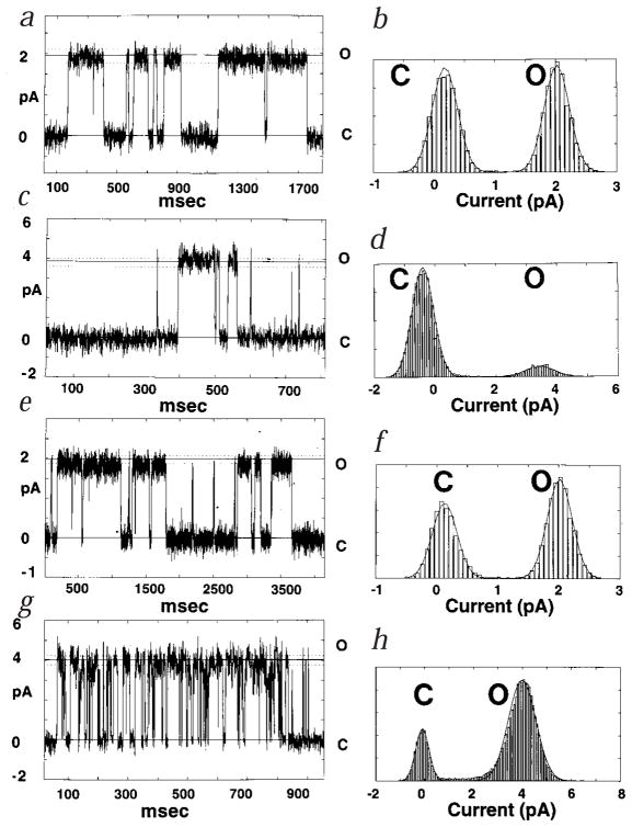
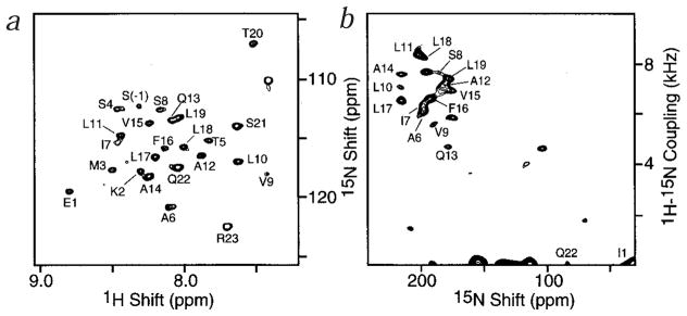
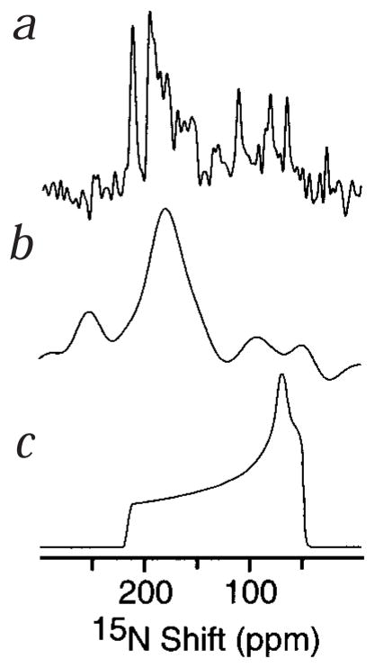
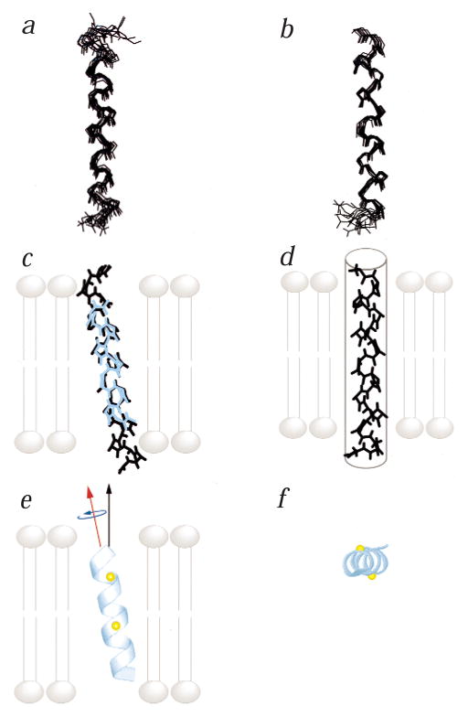
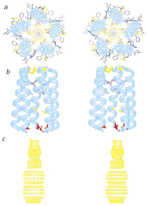
The structures of functional peptides corresponding to the predicted channel-lining M2 segments of the nicotinic acetylcholine receptor (AChR) and of a glutamate receptor of the NMDA subtype (NMDAR) were determined using solution NMR experiments on micelle samples, and solid-state NMR experiments on bilayer samples. Both M2 segments form straight transmembrane alpha-helices with no kinks. The AChR M2 peptide inserts in the lipid bilayer at an angle of 12 degrees relative to the bilayer normal, with a rotation about the helix long axis such that the polar residues face the N-terminal side of the membrane, which is assigned to be intracellular. A model built from these solid-state NMR data, and assuming a symmetric pentameric arrangement of M2 helices, results in a funnel-like architecture for the channel, with the wide opening on the N-terminal intracellular side.


Figures

Fig. 1
Fig. 1
ah, Single-channel recordings from recombinant M2 peptides in lipid bilayers. The currents were recorded at constant voltage in symmetric 500 mM NaCl or KCl supplemented with 1 mM CaCl2, 5 mM HEPES pH 7.4. The currents of closed (C) and open (O) states are indicated by the solid lines. Dotted lines define the range set to discriminate the transitions between states, based on signal-to-noise measurements. The AChR M2 traces show bursts of channel activity with single-channel conductances of 37 ± 2 pS in 500 mM NaCl at 50 mV (a), and 38 ± 2 pS in 500 mM KCl at 100 mV (c). For NMDAR M2, single-channel conductances of 20 ± 2 pS NaCl (e), and 40 ± 3 pS in 500 mM KCl (g) were measured. Both NMDAR traces were recorded at 100 mV. In the corresponding histograms for AChR M2 (b, d) and for NMDAR M2 (f, h), the Gaussian fits of the data in NaCl (b, f) or KCl (d, h) indicate the respective probabilities of the open- (O) versus closed- (C) channel states.
Fig. 2
Fig. 2
Solution and solid-state 2D NMR spectra of AchR M2 peptides. a, Solution 1H chemical-shift/15N chemical-shift correlation HMQC NMR spectrum of AChR M2 in DPC micelles, at 20 °C. Four transients were acquired for each of 256 t1 increments. b, Solid-state 1H-15N dipolar/15N chemical-shift correlation PISEMA NMR spectrum of AChR M2 in oriented DMPC bilayers, at 22 °C. A total of 256 transients were acquired for each of 64 t1 values incremented by 40.8 μs.
Fig. 3
Fig. 3
One-dimensional solid-state 15N chemical-shift NMR spectra of M2 peptides in oriented lipid bilayers. a, Uniformly 15N-labeled AChR M2 in DMPC bilayers. b, Uniformly 15N-labeled NMDAR M2 in POPC/DOTAP bilayers. c, Simulated chemical-shift powder pattern of an immobile 15N amide site.
Fig. 4
Fig. 4
Superposition of the backbone heavy atoms for the 10 lowest energy structures of a, AChR M2 and b, NMDAR M2, determined by solution NMR in DPC micelles. The four residues preceding the native sequence of NMDA M2 were not constrained in the structure calculations and are not shown. c, Superposition of the average structure of the AChR M2 calculated from the solution NMR distance constraints (black), and the average structure determined from the solid-state NMR orientational constraints (cyan). Both structures are shown in the bilayer membrane, in the exact overall orientation determined from solid-state NMR. d, Average structure of NMDAR M2, calculated from solution NMR experiments. The peptide is shown in the transmembrane orientation (gray tube) determined from the one-dimensional solid-state NMR spectrum. The exact tilt in the membrane is not determined because of the limited 15N chemical-shift data for this peptide. e,f, Side (e) and top (f) views of the average structure of AChR M2 in lipid bilayers, determined from solid-state NMR orientational constraints. The peptide is shown in its exact overall orientation within the lipid bilayer. The N-terminus is on top in (e), and in front in (f). The Cα atoms of Ser 8 and Gln 13 are highlighted in yellow. The helix long axis (red arrow) is tilted 12° from the membrane normal (black arrow). The helix rotation about its long axis (blue arrow) is such that the polar residues Ser 8 and Gln 13 face the N-terminal side of the lipid bilayer. In (c), (d) and (e) the lipid bilayer membrane is shown in gray.
Fig. 5
Fig. 5
ac, Top (a) and side (b,c) stereo views of a model of the AChR M2 funnel-like pentameric bundle. The channel architecture was calculated using the solid-state NMR three-dimensional coordinates of the M2 helix in the lipid bilayer, and by imposing a symmetric pentameric organization. The top view has the C-terminal synaptic side in front. The wide mouth of the funnel is on the N-terminal, intracellular side of the pore. A sodium ion is confined within the pore. The side views have the C-terminus on top. In (b), only the side chains of the pore-lining residues, Glu 1, Ser 8, Val 15, Leu 18 and Gln 22, are shown. In (c), the dotted contour depicts the pore profile calculated using the program HOLE; it represents the minimum radial distance between the center of the pore lumen and the van der Waals protein contact. The hole radii for the residues facing the pore lumen are 3.3 Å (Glu 1); 4.3 Å (Ser 8); 2.9 Å (Val 15); 2.1 Å (Leu 18); 1.5 Å (Gln 22). Ribbon diagrams were drawn using MOLSCRIPT. The α– carbon backbone is shown in cyan; acidic residues are in red, basic residues in blue, polar residues in yellow, and lipophilic residues in purple.

References

    1. Colquhoun D, Sakmann B. From muscle endplate to brain synapses: a short history of synapses and agonist-activated ion channels. Neuron. 1998;20:381–387. - PubMed
    1. Lena C, Changeux JP. Pathological mutations of nicotinic receptors and nicotine-based therapies for brain disorders. Curr Opin Neurobiol. 1997;7:674–682. - PubMed
    1. Hollmann M, Heinemann S. Cloned glutamate receptors. Annu Rev Neurosci. 1994;17:31–108. - PubMed
    1. Unwin N. Acetylcholine receptor channel imaged in the open state. Nature. 1995;373:37–43. - PubMed
    1. Oiki S, Madison V, Montal M. Bundles of amphipathic transmembrane α-helices as a structural motif for ion-conducting channel proteins: studies on sodium channels and acetylcholine receptors. Proteins. 1990;8:226–236. - PubMed

Publication types

MeSH terms

LinkOut - more resources