. 1999 Mar;126(5):1139-46.
doi: 10.1038/sj.bjp.0702422.

Protective effect of cyclosporin A and FK506 from nitric oxide-dependent apoptosis in activated macrophages

Affiliations

Protective effect of cyclosporin A and FK506 from nitric oxide-dependent apoptosis in activated macrophages

S Hortelano et al. Br J Pharmacol. 1999 Mar.

Abstract

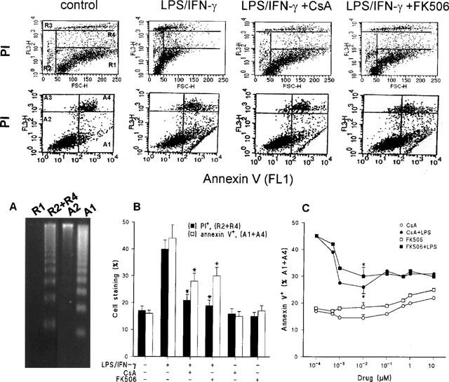
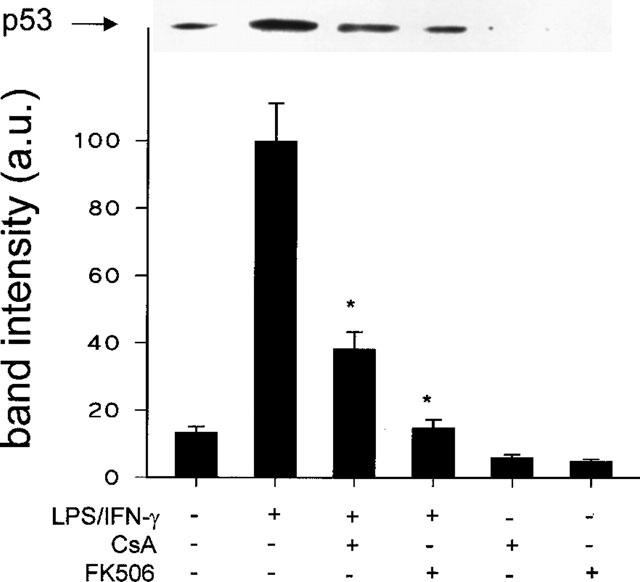
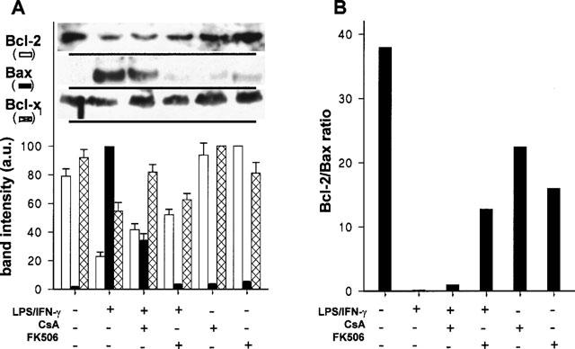
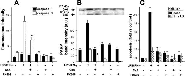
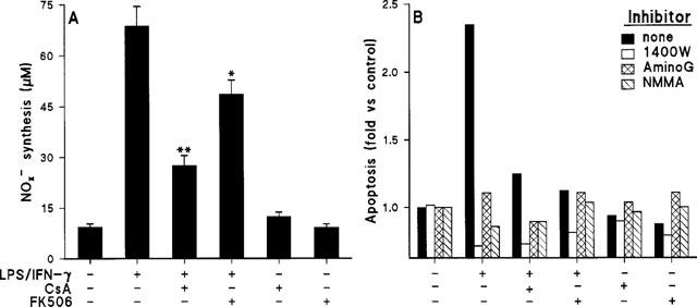
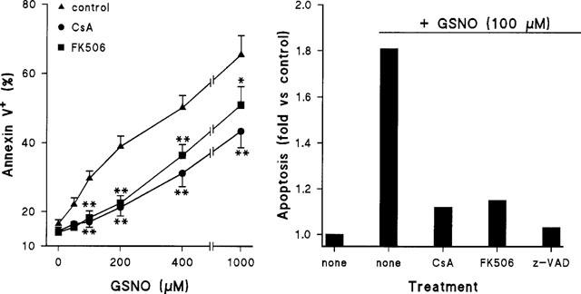
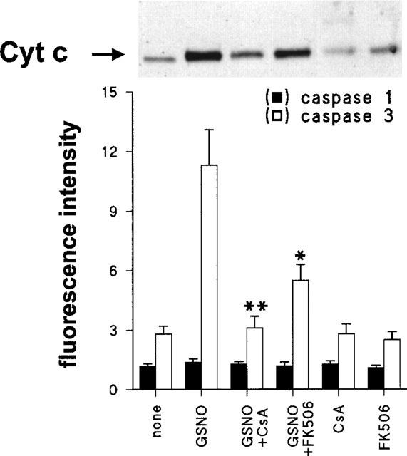
1. Activation of macrophages with lipopolysaccharide (LPS) and low doses of interferon-gamma (IFN-gamma) induced apoptotic death through a nitric oxide-dependent pathway. 2. Treatment of cells with the immunosuppressors cyclosporin A (CsA) or FK506 inhibited the activation-dependent apoptosis. 3. These drugs decreased the up-regulation of p53 and Bax characteristic of activated macrophages. Moreover, incubation of activated macrophages with CsA and FK506 contributed to maintain higher levels of Bcl-2 than in LPS/IFN-gamma treated cells. 4. The inhibition of apoptosis exerted by CsA and FK506 in macrophages was also observed when cell death was induced by treatment with chemical nitric oxide donors. 5. Incubation of macrophages with LPS/IFN-gamma barely affected caspase-1 but promoted an important activation of caspase-3. Both CsA and FK506 inhibited pathways leading to caspase-3 activation. Moreover, the cleavage of poly(ADP-ribose) polymerase, a well established caspase substrate, was reduced by these immunosuppressive drugs. 6. CsA and FK506 reduced the release of cytochrome c to the cytosol and the activation of caspase-3 in cells treated with nitric oxide donors. 7. These results indicate that CsA and FK506 protect macrophages from nitric oxide-dependent apoptosis and suggest a contribution of the macrophage to innate immunity under conditions of immunosuppression of the host.


Figures

Figure 1
Figure 1
CsA and FK506 decreased the amount of apoptotic macrophages and the binding of annexin V upon treatment with LPS/IFN-γ. Cultured peritoneal macrophages (106 cells) were stimulated for 24 h with combinations of 200 ng ml−1 of LPS and 10 u ml−1 of IFN-γ, CsA 10 nM or FK506 10 nM. The cells were analysed by flow cell cytometry after labelling with PI or staining with annexin V for the detection of the exposure of phosphatidylserine residues. LPS/IFN-γ activated macrophages corresponding to the R1, R2+R4, A2 and A1 panels were sorted (106 cells) and the DNA integrity was evaluated in agarose gels (A). The percentage of apoptotic cells (R2+R4 quadrants) after PI labelling (upper panels) or annexin V+ (A1+A4 quadrants of the lower panels) was quantified (B). The dose dependent effect of CsA or FK506 on the exposure of phosphatidylserine residues by control (open symbols) or activated macrophages (solid symbols) is shown in panel C. Results shown the mean±s.e.mean of three experiments. *P<0.01 with respect to the LPS/IFN-γ condition.
Figure 2
Figure 2
p53 up-regulation is inhibited in activated macrophages incubated with CsA or FK506. Cells were incubated with combinations of LPS (200 ng ml−1), IFN-γ (10 u ml−1), CsA (10 nM), or FK506 (10 nM) for 24 h. Total cell extracts were prepared and equal amounts of protein were analysed by Western blot using an anti-p53 Ab. Results show the p53 band corresponding to a representative experiment and the mean±s.e.mean of the densitometries of the bands from four experiments. *P<0.01 with respect to the LPS/IFN-γ condition.
Figure 3
Figure 3
CsA and FK506 attenuate the changes in the levels of proteins of the Bcl-2 family induced by treatment of macrophages with LPS/IFN-γ. Cells were treated as indicated in Figure 2 and soluble protein from the cell extracts was analysed by Western blot using anti-Bcl-2, anti-Bax and anti-Bcl-xl antibodies. Results show the densitometry of the bands (mean±s.e.mean) from four experiments (A). The ratio between the Bcl-2 and Bax levels corresponding to each condition is shown in B.
Figure 4
Figure 4
Effect of CsA and FK506 on LPS/IFN-γ-dependent caspase activation and PARP degradation. Macrophages were stimulated for 24 h with LPS/IFN-γ, CsA 10 nM, FK506 10 nM or combinations of these. Cell extracts were prepared and the activity of caspases-1 and -3 was measured using specific substrates and inhibitors (A). The extent of PARP cleavage, as an in vivo caspase substrate, was analysed by Western blot using a specific Ab (B). The effect of the caspase inhibitor z-VAD (20 μM) on apoptosis (propidium iodide staining) was evaluated in activated cells (C). Results (mean±s.e.mean, n=3) show the caspase activity (*P<0.01 vs the LPS/IFN-γ condition), the band intensities of PARP fragments, and the apoptosis upon normalization with respect to unstimulated cells.
Figure 5
Figure 5
Effect of CsA and FK506 on NO synthesis in activated macrophages. Protection from apoptosis in the absence of NO synthesis. Cells were treated as indicated in Figure 2 and the amount of nitrite plus nitrate released was measured (A). NOS activity was inhibited with 200 μM of 1400W, aminoguanidine (AminoG) or NMMA. Apoptosis was determined by staining the cells with propidium iodide and measuring the percentage of cells in the R2+R4 regions. Apoptosis (mean of two experiments) was expressed upon normalization with respect to control unstimulated cells (B). *P<0.05, **P<0.01 with respect to the LPS/IFN-γ condition.
Figure 6
Figure 6
CsA and FK506 inhibit the apoptosis induced by low doses of GSNO. Macrophages were treated for 6 h with the indicated doses of a fresh solution of GSNO and 10 nM of CsA or FK506. The percentage of annexin V+ cells was determined by flow cytometry (A). The protection exerted by z-VAD (20 μM) in cells treated with 100 μM GSNO was also measured (B). Results show the mean±s.e.mean (n=4) (A) or the mean of two experiments (B). *P<0.05, **P<0.005 with respect to the condition in the absence of immunomodulator.
Figure 7
Figure 7
Cytochrome c release and caspase-3 activation in macrophages treated with GSNO. Cells were incubated for 4 h with GSNO 200 μM, CsA 10 nM, FK506 10 nM or combinations of these. Cells were homogenized in conditions to preserve mitochondrial integrity and the cytosolic extracts were analysed to determine the amount of cytochrome c by Western blotting (17 kDa band), and the activity of caspase-1 and -3, respectively. Results show that the mean±s.e.mean of three experiments. *P<0.01, **P<0.001 with respect to cells treated with GSNO.

References

    1. AGARWAL M.L., TAYLOR W.R., CHERNOV M.V., CHERNOVA O.B., STARK G.R. The p53 network. J. Biol. Chem. 1998;273:1–4. - PubMed
    1. ALBINA J.E., CUI S., MATEO R.B., REICHNER J.S. Nitric oxide-mediated apoptosis in murine peritoneal macrophages. J. Immunol. 1993;150:5080–5085. - PubMed
    1. CALMELS S., HAINAUT P., OHSHIMA H. Nitric oxide induces conformational and functional modifications of wild-type p53 tumor suppressor protein. Cancer Res. 1997;57:3365–3369. - PubMed
    1. CARROLL M.C., PRODEUS A.P. Linkages of innate and adaptive immunity. Curr. Opin. Immunol. 1998;10:36–40. - PubMed
    1. CONDE M., ANDRADE J., BEDOYA F.J., SANTA MARIA C., SOBRINO F. Inhibitory effect of cyclosporin A and FK506 on nitric oxide production by cultured macrophages. Evidence of a direct effect of nitric oxide synthase activity. Immunology. 1995;84:476–481. - PMC - PubMed

Publication types

MeSH terms