. 1999 May;19(5):3423-34.
doi: 10.1128/MCB.19.5.3423.

Cellular activation triggered by the autosomal dominant polycystic kidney disease gene product PKD2

Affiliations

Cellular activation triggered by the autosomal dominant polycystic kidney disease gene product PKD2

T Arnould et al. Mol Cell Biol. 1999 May.

Abstract

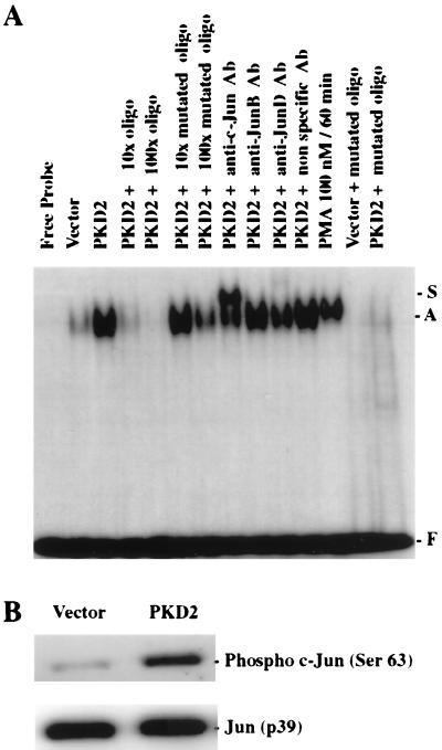
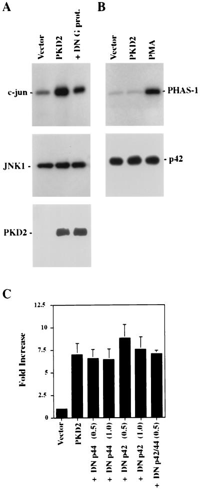
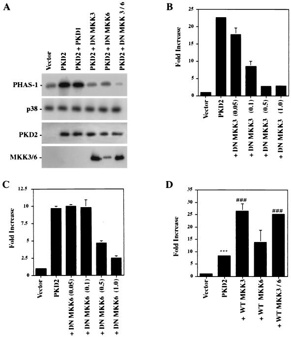
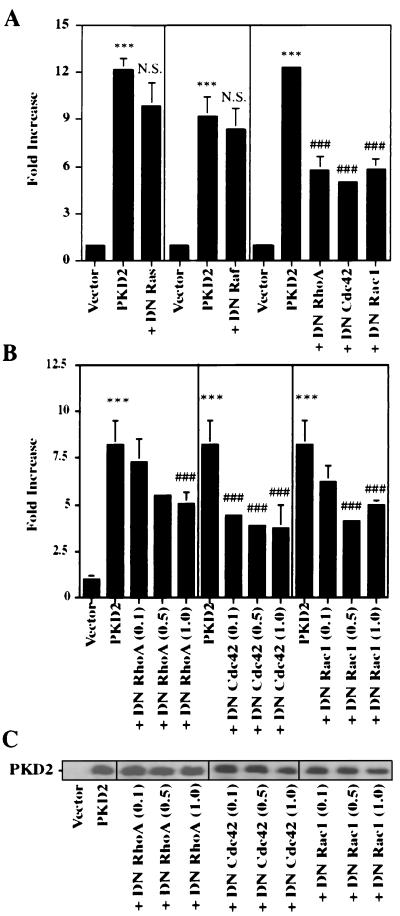
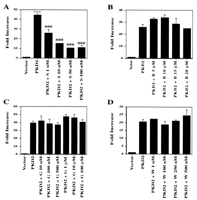
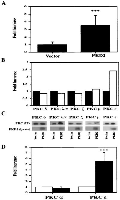
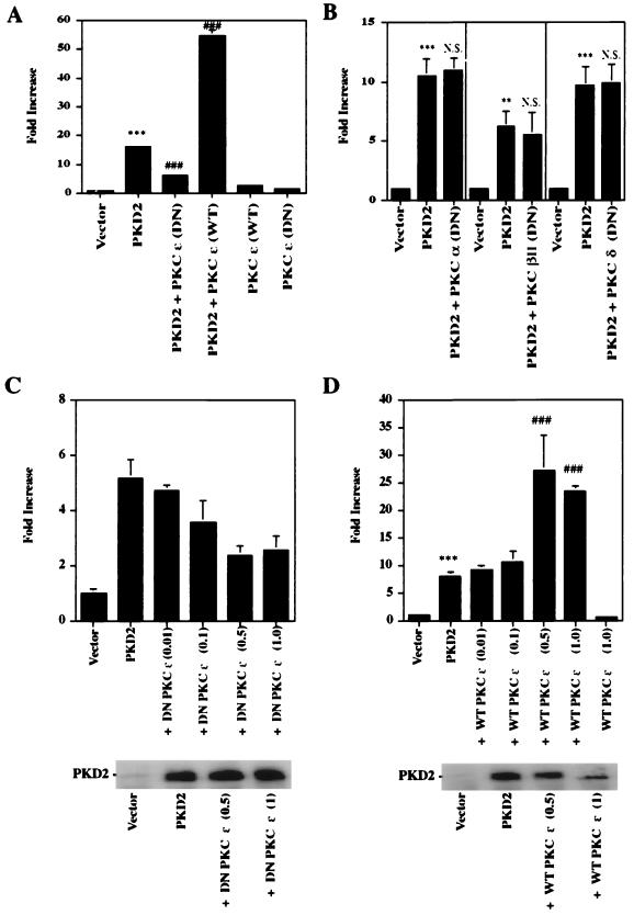
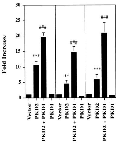
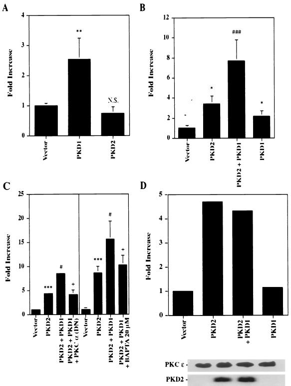
Autosomal dominant polycystic kidney disease (ADPKD) is caused by germ line mutations in at least three ADPKD genes. Two recently isolated ADPKD genes, PKD1 and PKD2, encode integral membrane proteins of unknown function. We found that PKD2 upregulated AP-1-dependent transcription in human embryonic kidney 293T cells. The PKD2-mediated AP-1 activity was dependent upon activation of the mitogen-activated protein kinases p38 and JNK1 and protein kinase C (PKC) epsilon, a calcium-independent PKC isozyme. Staurosporine, but not the calcium chelator BAPTA [1,2-bis(o-aminophenoxy)ethane-N,N,N', N'-tetraacetate], inhibited PKD2-mediated signaling, consistent with the involvement of a calcium-independent PKC isozyme. Coexpression of PKD2 with the interacting C terminus of PKD1 dramatically augmented PKD2-mediated AP-1 activation. The synergistic signaling between PKD1 and PKD2 involved the activation of two distinct PKC isozymes, PKC alpha and PKC epsilon, respectively. Our findings are consistent with others that support a functional connection between PKD1 and PKD2 involving multiple signaling pathways that converge to induce AP-1 activity, a transcription factor that regulates different cellular programs such as proliferation, differentiation, and apoptosis. Activation of these signaling cascades may promote the full maturation of developing tubular epithelial cells, while inactivation of these signaling cascades may impair terminal differentiation and facilitate the development of renal tubular cysts.


Figures

FIG. 1
FIG. 1
PKD2 activates AP-1- and c-Jun (TRE)-dependent transcription. (A) PKD2 triggers activation of an AP-1 reporter construct containing four tandem AP-1-binding sites and a c-Jun reporter construct containing three repeats of the second TRE of the c-Jun promoter (Jun2TRE). HEK 293T cells were transfected with 1 μg of vector (CDM8) (white bars) or PKD2 (black bars), as well as luciferase reporter constructs for AP-1, c-Jun, c-myc, CREB, NF-κB, or c-Fos. Transactivation was determined after 36 h of incubation, and luciferase activity was expressed after normalization for β-galactosidase activity as fold increase over the vector control. The results represent the means ± SD of transfections performed in triplicate. (B) PKD2 activates the AP-1 reporter in a dose-dependent fashion. Increasing amounts of PKD2 vector were transfected to transactivate the AP-1 reporter construct. The total amount of plasmid DNA (2 μg/transfection) was balanced with vector (CDM8). (C) PKD2 activates the Jun2TRE reporter in a dose-dependent fashion. Increasing amounts of PKD2 vector were transfected to transactivate the Jun2TRE reporter. The total amount of plasmid DNA (2 μg/transfection) was balanced with vector (CDM8). (D) PKD2 activates the collagenase promoter. HEK 293T cells were cotransfected with 1 μg of PKD2 or vector (CDM8) and a collagenase promoter construct containing only a single AP-1-binding site. Cells were stimulated with 10% serum for 8 h. The results represent the means ± SD of transfections performed in triplicate. ∗, P < 0.05; ∗∗, P < 0.01; ∗∗∗, P < 0.001.
FIG. 2
FIG. 2
PKD2 triggers AP-1-binding activity. (A) PKD2 induces binding of nuclear proteins to the AP-1/TRE DNA-binding site. Nuclear extracts were isolated from HEK 293T cells transfected with either vector or PKD2 or from cells stimulated with 100 nM PMA for 60 min and analyzed by gel retardation assay for AP-1 binding. No nuclear extract was added to the radiolabeled AP-1 oligonucleotide in lane 1 (Free Probe). Addition of a 10- or 100-fold excess of unlabeled probe prevented the formation of a DNA-protein complex, while addition of a 10- or 100-fold excess of mutated AP-1 oligonucleotide had little effect. A significant supershift of the DNA-protein complex was observed after the addition of a monoclonal antibody to c-Jun but not an antibody to JunB or JunD or a nonspecific antibody. A mutated AP-1 oligonucleotide failed to bind nuclear proteins isolated from PKD2-transfected HEK 293T cells. The positions of the AP-1 complex (A), supershift (S), and free probe (F) are indicated. (B) Western blot analysis showing the phosphorylation of c-Jun after cotransfection of a His-tagged c-Jun plasmid with a construct encoding PKD2 or a vector control (CDM8). c-Jun phosphorylated on serine 63 was detected with a specific polyclonal antiserum. To demonstrate equal amounts of total c-Jun, the membrane was reprobed with an anti-Jun antiserum.
FIG. 3
FIG. 3
PKD2 activates the JNK1 but not the MAPK p42. (A) HEK 293T cells were cotransfected with HA-tagged JNK1 and vector control (CDM8), PKD2, or PKD2 in combination with dominant-negative (DN) mutants of the small G proteins Cdc42, Rac1, and RhoA at equal ratios. Cells were harvested after 24 h, and immunoprecipitated JNK1 was incubated with GST-c-Jun(1–79) in the presence of [γ-32P]ATP. Incorporated radioactivity was visualized by SDS–10% PAGE and autoradiography. PKD2 increases JNK1 activity (top panel); this activation was inhibited by the DN Cdc42, Rac1, and RhoA. Western blot analysis revealed equal amounts of precipitated kinases (middle panel); the expression of PKD2 was not affected by the presence of the DN Cdc42, Rac1, and RhoA (bottom panel). (B) HEK 293T cells were cotransfected with HA-tagged p42 in combination with a vector control (CDM8) or PKD2 at equal ratios. Cells were harvested after 24 h, and immunoprecipitated p42 was incubated with PHAS-1 in the presence of [γ-32P]ATP. Incorporated radioactivity was visualized by SDS–12% PAGE and autoradiography. Only stimulation with 50 nM PMA for 30 min resulted in activation of p42. The lower panel demonstrates equal amounts of p42 kinase in each condition as determined by Western blot analysis. (C) PKD2-mediated AP-1 activation is unaffected by DN mutants of the MAPK p42 or p44. HEK 293T cells were transfected with the AP-1 reporter construct, vector control, PKD2, or PKD2 in combination with DN mutants of p42 and p44. Transactivation was determined after 36 h of incubation, and luciferase activity was expressed after normalization for β-galactosidase activity as fold increase over the vector control. The results represent the means ± SD of transfections performed in triplicate.
FIG. 4
FIG. 4
PKD2 activates p38 MAPK. (A) Dominant-negative (DN) mutants of MKK3 and MKK6 significantly inhibit PKD2-mediated p38 activation. In vitro kinase assays were performed after transfection of HEK 293T cells with HA-tagged p38 and PKD2 or a vector control (CDM8). Immunoprecipitated kinases were incubated with PHAS-1 as an exogenous substrate in the presence of [γ-32P]ATP. Incorporated radioactivity was visualized by SDS–12% PAGE and autoradiography. The amounts of immunoprecipitated p38, the expression of PKD2, and the expression of the DN MKK3 and MKK6 were monitored by Western blot analysis. PKD2 activates p38 in an MKK3- and MKK6-dependent fashion; coexpression of C-terminal PKD1 does not contribute to the PKD2-mediated p38 activation. (B) A DN mutant of MKK3 significantly inhibits PKD2-mediated AP-1 activation in a dose-dependent fashion. AP-1 luciferase assays were performed after transfection of HEK 293T cells with 1 μg of PKD2 and increasing amounts of a DN mutant of MKK3. The results represent the means ± SD of transfections performed in triplicate. (C) A DN mutant of MKK6 significantly inhibits PKD2-mediated AP-1 activation in a dose-dependent fashion. AP-1 luciferase assays were performed after transfection of HEK 293T cells with 1 μg of PKD2 and increasing amounts of a DN mutant of MKK6. The results represent the means ± SD of transfections performed in triplicate. (D) Wild-type (WT) MKK3 and MKK6 enhance PKD2-mediated AP-1 activation. AP-1 luciferase assays were performed after transfection of HEK 293T cells with 1 μg of PKD2 in combination with WT MKK3 or MKK6. The results represent the means ± SD of transfections performed in triplicate. ∗∗∗, significantly different from control vector (P < 0.001); ⋕⋕⋕⋕, significantly different from PKD2-transfected cells (P < 0.001).
FIG. 5
FIG. 5
Dominant-negative (DN) mutants of the GTP-binding proteins RhoA, Cdc42, and Rac1 inhibit PKD2-mediated AP-1 activation. (A) DN mutants of RhoA, Cdc42, and Rac1 but not Ras or Raf-1 blocked AP-1 activation in HEK 293T cells transfected with PKD2. Results represent the means ± SD of three independent experiments. (B) DN mutants of RhoA, Cdc42, and Rac1 inhibit PKD2-mediated AP-1 activation in a dose-dependent fashion. Results represent the means ± SD of three independent experiments. (C) DN mutants of RhoA, Cdc42, and Rac1 do not affect the expression of PKD2. PKD2 expression, monitored by Western blot analysis, was unaffected by increasing amounts of cotransfected DN RhoA, Cdc42, or Rac1. ∗∗∗, significantly different from control (P < 0.001); ⋕⋕⋕, significantly different from PKD2-transfected cells (P < 0.001); N.S., not significant.
FIG. 6
FIG. 6
Effects of staurosporine (S), BAPTA-AM (B), wortmannin (W), and genistein (G) on PKD2-mediated AP-1 activation. HEK 293T cells were transiently cotransfected with PKD2 or a vector control (CDM8) and were exposed to increasing concentrations of staurosporine (A), BAPTA (B), genistein (C), and wortmannin (D), added for the last 6 h of the incubation period. Transactivation of the AP-1 reporter construct was determined after 36 h of incubation, and luciferase activity was expressed as fold increase over the vector control after normalization for β-galactosidase activity. Representative results of two experiments, performed in triplicate, are expressed as means ± SD. ∗∗∗, significantly different from control vector (P < 0.001); ⋕⋕⋕, significantly different from PKD2-transfected cells (P < 0.001).
FIG. 7
FIG. 7
PKD2 activates PKC ɛ. (A) PKD2 increases total PKC activity in HEK 293T cells. HEK 293T cells were transiently transfected with a vector encoding PKD2 or a vector control, and total PKC activity was measured by a colorimetric assay. Results are expressed as fold increase over the vector control for six independent experiments. (B) PKD2 activates PKC ɛ but not other calcium-independent PKC isozymes. HEK 293T cells were transiently transfected with PKD2 (black bars) or a vector control (CDM8) (white bars). The different PKC isozymes (PKC δ, λ/τ, ζ, μ, and ɛ) were immunoprecipitated with specific monoclonal antibodies and analyzed by in vitro kinase assays. (C) The amounts of immunoprecipitated PKC isozymes (IP) and expression of PKD2 were monitored by Western blot analysis. (D) PKD2 activates PKC ɛ but not PKC α. The differential activation of PKC α and ɛ by PKD2 was confirmed by in vitro kinase assays (three independent experiments) and expressed as fold increase over the vector control. ∗∗∗, P < 0.001.
FIG. 8
FIG. 8
PKD2-mediated AP-1 activation requires PKC ɛ. (A) A dominant-negative (DN) PKC ɛ mutant blocks PKD2-mediated AP-1 activation. HEK 293T cells were transiently cotransfected with a vector control (CDM8) or PKD2 and a DN mutant of PKC ɛ or wild-type (WT) PKC ɛ at a ratio of 2:1. Transfections with 1 μg of the WT and DN PKC ɛ constructs alone are also shown. The experiment was performed in triplicate. (B) DN PKC α, βII, and δ mutants do not affect PKD2-mediated AP-1 activity. HEK 293T cells were transiently cotransfected with a vector control (CDM8) or PKD2, together with a DN mutant of PKC α, βII, or δ at a ratio of 2:1. The experiment was performed in triplicate. (C) A DN PKC ɛ mutant inhibits PKD2-mediated AP-1 activation in a dose-dependent fashion but does not affect PKD2 expression. HEK 293T cells were transiently cotransfected with a vector control (CDM8) or PKD2 and with increasing amounts of a DN mutant of PKC ɛ. PKD2 expression was monitored by Western blot analysis. (D) Wild-type (WT) PKC ɛ augments PKD2-mediated AP-1 activation. HEK 293T cells were transiently cotransfected with a vector control (CDM8) or PKD2 and with increasing amounts of WT PKC ɛ. PKD2 expression was monitored by Western blot analysis. ∗∗, significantly different from vector control (P < 0.01); ∗∗∗, significantly different from control vector (P < 0.001); ⋕⋕⋕, significantly different from PKD2-transfected cells (P < 0.001); N.S., not significantly different from PKD2-transfected cells.
FIG. 9
FIG. 9
PKD1 augments PKD2-mediated AP-1 activation. HEK 293T cells were transfected with vector control (CDM8), PKD2 together with CD16.7 (or CD16.7 fused to the last 112 amino acids of the C-terminal domain of PKD1), or PKD1 alone. The means ± SD of three independent experiments performed in triplicate are shown. ∗∗, significantly different from control vector (P < 0.01); ∗∗∗, significantly different from control vector (P < 0.001); ⋕⋕⋕, significantly different from PKD2-transfected cells (P < 0.001).
FIG. 10
FIG. 10
PKD1-mediated augmentation of PKD2 activities requires PKC α. (A) The C-terminal domain of PKD1 but not PKD2 activates PKC α. The PKC α isozyme was immunoprecipitated from PKD1, PKD2, or vector control-transfected HEK 293T cells; the activity was determined by in vitro kinase assays. The means ± SD of three experiments were expressed as fold increase over the vector control. (B) PKD1 increases the total PKC activity triggered by PKD2. HEK 293T cells were transfected with PKD1, PKD2, or a vector control (CDM8). The means ± SD of the total PKC activity of six independent experiments were expressed as fold increase over the vector control. (C) A dominant-negative (DN) PKC α mutant and the calcium chelator BAPTA block the effect of PKD1 upon PKD2-mediated AP-1 activation. HEK 293T cells were transiently cotransfected with a vector control (CDM8), PKD1, and PKD2 in combination with a DN mutant of PKC α. In one experiment, BAPTA-AM (20 μM) was added for 6 h. Experiments were performed in triplicate. (D) PKD1 has no effect on the PKD2-mediated activation of PKC ɛ. The PKC ɛ isozyme was immunoprecipitated from HEK 293T cells transfected with vector control, PKD1, or PKD2. The PKC ɛ activity was determined by in vitro kinase assays. Immunoprecipitates of PKC ɛ and PKD2 expression were monitored by Western blot analysis. ∗, significantly different from vector control (P < 0.05); ∗∗, significantly different from vector control (P < 0.01); ∗∗∗, significantly different from control vector (P < 0.001); ⋕, significantly different from PKD2-transfected cells (P < 0.05); ⋕⋕⋕, significantly different from PKD2-transfected cells, P < 0.05; +, significantly different from PKD1- and PKD2-transfected cells (P < 0.05); N.S., not significantly different from control cells.

References

    1. Arnould T, Kim E, Tsiokas L, Jochimsen F, Gruning W, Chang J D, Walz G. The polycystic kidney disease 1 gene product mediates protein kinase C alpha-dependent and c-Jun N-terminal kinase-dependent activation of the transcription factor AP-1. J Biol Chem. 1998;273:6013–6018. - PubMed
    1. Bagrodia S, Derijard B, Davis R J, Cerione R A. Cdc42 and PAK-mediated signaling leads to Jun kinase and p38 mitogen-activated protein kinase activation. J Biol Chem. 1995;270:27995–27998. - PubMed
    1. Boyle W J, Smeal T, Defize L H, Angel P, Woodgett J R, Karin M, Hunter T. Activation of protein kinase C decreases phosphorylation of c-Jun at sites that negatively regulate its DNA-binding activity. Cell. 1991;64:573–584. - PubMed
    1. Chen Y, Takeshita A, Ozaki K, Kitano S, Hanazawa S. Transcriptional regulation by transforming growth factor beta of the expression of retinoic acid and retinoid X receptor genes in osteoblastic cells is mediated through AP-1. J Biol Chem. 1996;271:31602–31606. - PubMed
    1. Coso O A, Chiariello M, Yu J C, Teramoto H, Crespo P, Xu N, Miki T, Gutkind J S. The small GTP-binding proteins Rac1 and Cdc42 regulate the activity of the JNK/SAPK signaling pathway. Cell. 1995;81:1137–1146. - PubMed

Publication types

MeSH terms

Substances