. 1999 May;19(5):3435-42.
doi: 10.1128/MCB.19.5.3435.

Prohibitins regulate membrane protein degradation by the m-AAA protease in mitochondria

Affiliations

Prohibitins regulate membrane protein degradation by the m-AAA protease in mitochondria

G Steglich et al. Mol Cell Biol. 1999 May.

Abstract

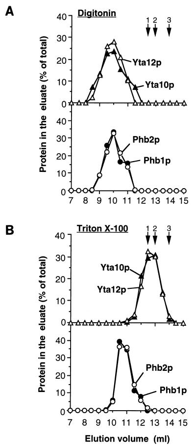
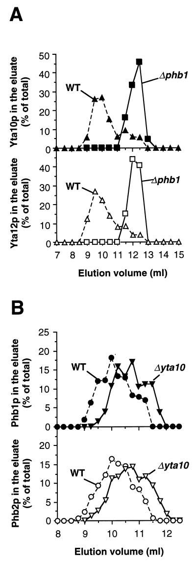
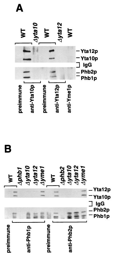
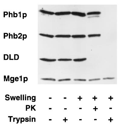
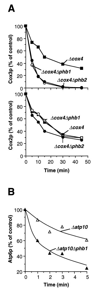
Prohibitins comprise a protein family in eukaryotic cells with potential roles in senescence and tumor suppression. Phb1p and Phb2p, members of the prohibitin family in Saccharomyces cerevisiae, have been implicated in the regulation of the replicative life span of the cells and in the maintenance of mitochondrial morphology. The functional activities of these proteins, however, have not been elucidated. We demonstrate here that prohibitins regulate the turnover of membrane proteins by the m-AAA protease, a conserved ATP-dependent protease in the inner membrane of mitochondria. The m-AAA protease is composed of the homologous subunits Yta10p (Afg3p) and Yta12p (Rca1p). Deletion of PHB1 or PHB2 impairs growth of Deltayta10 or Deltayta12 cells but does not affect cell growth in the presence of the m-AAA protease. A prohibitin complex with a native molecular mass of approximately 2 MDa containing Phb1p and Phb2p forms a supercomplex with the m-AAA protease. Proteolysis of nonassembled inner membrane proteins by the m-AAA protease is accelerated in mitochondria lacking Phb1p or Phb2p, indicating a negative regulatory effect of prohibitins on m-AAA protease activity. These results functionally link members of two conserved protein families in eukaryotes to the degradation of membrane proteins in mitochondria.


Figures

FIG. 1
FIG. 1
Cofractionation of Phb1p and Phb2p with the m-AAA protease upon sizing chromatography. Wild-type mitochondria were solubilized in buffer A containing 1% digitonin (A) or 0.2% Triton X-100 (B) as described in Materials and Methods. Extracts were fractionated by Superose 6 chromatography. Eluate fractions (0.5 ml) were analyzed by SDS-PAGE and immunostaining with antisera directed against Yta10p (▴), Yta12p (▵), Phb1p (●), and Phb2p (○). Hsp60 (12.5 ml; arrow 1), thyroglobulin (13 ml; arrow 2), apoferritin (14 ml; arrow 3), alcohol dehydrogenase (15.5 ml; not shown) and bovine serum albumin (16.5 ml; not shown) were used for calibration.
FIG. 2
FIG. 2
Gel filtration analysis of mitochondrial extracts lacking prohibitins or m-AAA protease. (A) Wild-type (WT) and Δphb1 mitochondria or (B) wild-type (WT) and Δyta10 mitochondria were lysed in buffer A containing 1% digitonin and fractionated by sizing chromatography as described in Materials and Methods. In panel B, fractions of 0.25 ml were collected to increase the resolution. Eluate fractions were analyzed by SDS-PAGE and immunostaining with antisera directed against (A) Yta10p (▴ and ■) and Yta12p (▵ and □) or (B) Phb1p (● and ▾) and Phb2p (○ and ▿). Proteins used for calibration are described in the legend to Fig. 1.
FIG. 3
FIG. 3
Coimmunoprecipitation of the m-AAA protease with the Phb1p-Phb2p complex. Mitochondria isolated from the indicated strains were solubilized and subjected to immunoprecipitation as described in Materials and Methods, using antisera directed against Yta10p, Yta12p, and Yme1p (A) or Phb1p and Phb2p (B) and the respective preimmune sera. The immunoprecipitates were analyzed by SDS-PAGE and immunostaining. WT, wild type.
FIG. 4
FIG. 4
Topology of Phb1p and Phb2p in the inner membrane. Mitochondria were subfractionated by osmotic disruption of the outer membrane, and accessibility to externally added trypsin or proteinase K (PK) was examined as described in Materials and Methods. Mitochondrial fractions were analyzed by SDS-PAGE and immunostaining using antisera directed against Phb1p, Phb2p, d-lactate dehydrogenase (DLD; as a marker for the intermembrane space), and Mge1p (as a marker for the mitochondrial matrix).
FIG. 5
FIG. 5
Accelerated degradation of nonassembled inner membrane proteins by the m-AAA protease in the absence of prohibitins. Mitochondrial translation products were labeled in the strains indicated, and cells were further incubated at 30°C to assess the stability of newly synthesized polypeptides as described in Materials and Methods. (A) Proteolysis of Cox3p and Cox2p in Δcox4 cells. Rate constants for proteolysis of Cox3p: k = 0.044 min−1 (in Δcox4 cells), k = 0.13 min−1 (in Δcox4 Δphb1 cells), and k = 0.15 min−1 (in Δcox4 Δphb2 cells). Rate constants for proteolysis of Cox2p: k = 0.036 min−1 (in Δcox4 cells), k = 0.037 min−1 (in Δcox4 Δphb1 cells), and k = 0.033 min−1 (in Δcox4 Δphb2 cells). (B) Proteolysis of Atp6p by the m-AAA protease in Δatp10 cells. Rate constants for proteolysis of Atp6p: k = 0.12 min−1 (in Δatp10 cells) and k = 0.29 min−1 (in Δatp10 Δphb1 cells).
FIG. 6
FIG. 6
Impaired cell growth of Δphb1 and Δphb2 strains lacking m-AAA protease. PHB1 and PHB2 were disrupted in wild-type (WT), Δyta10, Δyta12, and Δyme1 cells by PCR-targeted homologous recombination (36). Disruptants were grown on YPD medium (rich medium containing 2% glucose) and harvested in exponential phase. Equal amounts of cells were spotted onto YPD agar plates and incubated at 30°C for 16 h. Slow growth of Δyta10 and Δyta12 cells lacking PHB1 or PHB2 was detected upon prolonged incubation of the plates (data not shown).
FIG. 7
FIG. 7
Sequence similarity of E. coli HflC with prohibitin family members. Amino acid sequences of S. cerevisiae Phb1p (ScPhb1p; P40961) and prohibitin from Arabidopsis thaliana (AtPhb; U69155) and Homo sapiens (hsPhb; P35232) were aligned with E. coli HflC (EcHflC; P25661) by using the Clustal W program, version 1.7. Identical amino acid residues are shaded in black. Sequence identity between prohibitins of different organisms is over 50%. Predicted transmembrane segments at the N termini are underlined. E. coli HflC show 20% identical and 33% homologous residues with S. cerevisiae Phb1p and 13% identical and 29% homologous residues with S. cerevisiae Phb2p. We observed a similar degree of similarity between E. coli HflK and S. cerevisiae Phb1p and Phb2p (data not shown).

References

    1. Ackermann S H, Tzagoloff A. ATP10, a yeast nuclear gene required for the assembly of the mitochondrial F1-F0 complex. J Biol Chem. 1990;265:9952–9959. - PubMed
    1. Arlt H, Steglich G, Perryman R, Guiard B, Neupert W, Langer T. The formation of respiratory chain complexes in mitochondria is under the proteolytic control of the m-AAA-protease. EMBO J. 1998;17:4837–4847. - PMC - PubMed
    1. Arlt H, Tauer R, Feldmann H, Neupert W, Langer T. The YTA10-12-complex, an AAA protease with chaperone-like activity in the inner membrane of mitochondria. Cell. 1996;85:875–885. - PubMed
    1. Berger K H, Yaffe M P. Prohibitin family members interact genetically with mitochondrial inheritance components in Saccharomyces cerevisiae. Mol Cell Biol. 1998;18:4043–4052. - PMC - PubMed
    1. Brodsky J L, McCracken A A. ER-associated and proteasome-mediated protein degradation: how two topologically restricted events came together. Trends Cell Biol. 1997;7:151–156. - PubMed

Publication types