. 1999 May;19(5):3600-6.
doi: 10.1128/MCB.19.5.3600.

Identification of the novel player deltaEF1 in estrogen transcriptional cascades

Affiliations

Identification of the novel player deltaEF1 in estrogen transcriptional cascades

E M Chamberlain et al. Mol Cell Biol. 1999 May.

Abstract

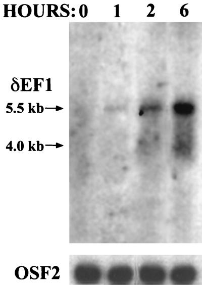
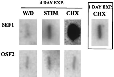
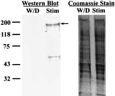
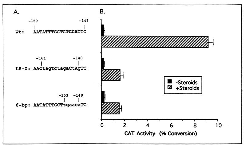
Although many genes are regulated by estrogen, very few have been shown to directly bind the estrogen receptor complex. Therefore, transcriptional cascades probably occur in which the estrogen receptor directly binds to a target gene that encodes another transcription factor that subsequently regulates additional genes. Through the use of a differential display assay, a transcription factor has been identified that may be involved in estrogen transcriptional cascades. This report demonstrates that transcription factor deltaEF1 is induced eightfold by estrogen in the chick oviduct. Furthermore, the regulation by estrogen occurs at the transcriptional level and is likely to be a direct effect of the estrogen receptor complex, as it does not require concomitant protein synthesis. A putative binding site was identified in the 5'-flanking region of the chick ovalbumin gene identifying it as a possible target gene for regulation by deltaEF1. Characterization of this binding site revealed that deltaEF1 binds to and regulates the chick ovalbumin gene. Thus, a novel regulatory cascade that is triggered by estrogen has been defined.


Figures

FIG. 1
FIG. 1
Differential display gel showing the time course of induction of oviduct cDNAs by estrogen. The arrow points to the δEF1 cDNA, which fits the criterion of increased abundance throughout the time points after 0.5 h. Each lane represents a sample from an individual chick. The values at the top are times of exposure to an intravenous injection of 17-β-estradiol.
FIG. 2
FIG. 2
δEF1 mRNA is induced quickly upon treatment with 17-β-estradiol. Poly(A)+ RNA (2 μg) was extracted from chick oviducts at the indicated times after injection with 17-β-estradiol and subjected to Northern blot analysis using a 0.25-kb cDNA fragment corresponding to nucleotides 2199 to 2449 of the δEF1 gene. This time course agrees well with the induction seen in Fig. 1. The lower panel is the same Northern blot probed with a 1.4-kb fragment of OSF2 cDNA, a gene that is not regulated by estrogen, to show equal loading among the lanes.
FIG. 3
FIG. 3
Regulation of δEF1 gene expression by estrogen is at the transcriptional level and does not require concomitant protein synthesis. Nuclei isolated from the oviducts of chicks that were either withdrawn from DES for 5 days (W/D), DES stimulated (STIM), or withdrawn from DES for 5 days and then injected with cycloheximide for 2.5 h and estrogen for 2 h (CHX) were used in a nuclear runon transcriptional assay. The blot reveals that δEF1 is regulated at the transcriptional level and does not require concomitant protein synthesis for expression. The main group of six panels shows the nuclear runon after 4 days of exposure (EXP.) to Hyperfilm (Amersham). The boxed panel on the right shows the effect of cycloheximide and estrogen after 1 day of exposure for clarity. OSF2 cDNA was used as a control.
FIG. 4
FIG. 4
δEF1 protein levels are also induced by estrogen. Chick nuclear protein was extracted from DES-stimulated chicks (Stim) or chicks from which DES had been withdrawn for 5 days (W/D). Fifty micrograms of nuclear protein was blotted with an antibody specific for δEF1. The arrow indicates the full-length δEF1 protein. The smaller band likely represents a proteolytic fragment, as inclusion of a protease inhibitor cocktail greatly reduced the intensity of this band (data not shown). The panel on the right shows Coomassie staining to demonstrate approximate equal loading of samples. The values on the left are molecular sizes in kilodaltons.
FIG. 5
FIG. 5
δEF1 is the component of oviduct nuclear extracts that binds to the ovalbumin gene. A gel mobility shift assay was performed by using the δEF1 binding site from the ovalbumin gene as a probe. Lane 1 shows the probe (−159 to −141) alone. The ovalbumin probe was incubated with 8 μg of oviduct nuclear protein extract (lanes 2 to 4) or with in vitro-transcribed-translated δEF1 protein (lanes 6 to 8) or a mock in vitro transcription-translation reaction mixture (lane 5). Addition of preimmune sera (lane 3) did not affect binding to the ovalbumin probe. Addition of 1 μl of δEF1 antibody (lane 4) abolished the complexes seen with the probe and oviduct nuclear protein (lane 2). Several smaller complexes were seen when δEF1 antibody was included in the binding reaction mixture and were likely due to other proteins binding to the probe in the absence of δEF1 binding. Incubation of the probe with 2 μl of a 50-μl δEF1 in vitro transcription-translation reaction mixture resulted in the formation of a complex (lane 6) that was the same size as complexes formed with nuclear protein extracts (compare lanes 2 and 6). Addition of the δEF1 antibody to the in vitro-transcribed-translated δEF1 abolished all binding (lane 8), while preimmune serum did not affect binding (lane 7). Lane 5 shows the ovalbumin probe with 2 μl of a mock in vitro transcription-translation reaction mixture added.
FIG. 6
FIG. 6
Mutation of the δEF1 site in the ovalbumin gene causes sixfold attenuation of activity. (A) Sequences of the ovalbumin gene in which mutations were made. Nucleotide positions relative to the transcription start site are indicated. The δEF1 site is indicated by boldface type and lowercase letters indicate mutant bases. (B) Transient transfection of constructs into chick primary tubular gland cells. Following transfection, the cells were cultured in the absence (black bars) or the presence (cross-hatched bars) of estrogen and glucocorticoid. Glucocorticoid is needed to achieve maximal activation of the ovalbumin promoter. Standard errors are indicated by bars. The graph depicted here is a composite of four experiments with each construct tested in duplicate for each treatment. Wt, wild type.
FIG. 7
FIG. 7
Overexpression of δEF1 in the absence of steroid hormones activates the ovalbumin gene. The δEF1 expression construct was transiently cotransfected into chick primary oviduct cells along with the wild-type ovalbumin reporter construct pOvCAT-.900. The level of induction was normalized to that seen in oviduct cells cotransfected with an empty expression vector in the absence of steroid hormones (−S). The amount of δEF1 expression plasmid transfected is shown with the total amount of DNA being held constant by the addition of an empty expression vector. Transfection was done in both the absence (−S) and the presence (+S) of estrogen and glucocorticoid. The graph shown is for a representative experiment, one of three such experiments, with each construct done in duplicate per treatment. The error bars represent the ranges of duplicate samples.

References

    1. Ashbruner M, Chihara C, Meltzer C, Richard G. Temporal control of puffing in polytene chromosomes. Cold Spring Harbor Symp Quant Biol. 1974;38:655–662. - PubMed
    1. Barrett C E. Postmenopausal estrogen and heart disease. Atherosclerosis. 1995;118:S7–S10. - PubMed
    1. Behl C, Widmann M, Trapp T, Holsboer F. 17-Beta estradiol protects neurons from oxidative stress-induced cell death in vitro. Biochem Biophys Res Commun. 1995;216:473–482. - PubMed
    1. Chiou S T, Chang W C. Insulin-like growth factor I stimulates transcription of the c-jun proto-oncogene in Balb/C 3T3 cells. Biochem Biophys Res Commun. 1992;183:524–531. - PubMed
    1. Dean D M, Jones P S, Sanders M M. Regulation of the chick ovalbumin gene by estrogen and corticosterone requires a novel DNA element that binds a labile protein, Chirp-1. Mol Cell Biol. 1996;16:2015–2024. - PMC - PubMed

Publication types

LinkOut - more resources