. 1999 May;19(5):3727-35.
doi: 10.1128/MCB.19.5.3727.

Distinct mechanisms of activation of Stat1 and Stat3 by platelet-derived growth factor receptor in a cell-free system

Affiliations

Distinct mechanisms of activation of Stat1 and Stat3 by platelet-derived growth factor receptor in a cell-free system

M L Vignais et al. Mol Cell Biol. 1999 May.

Abstract

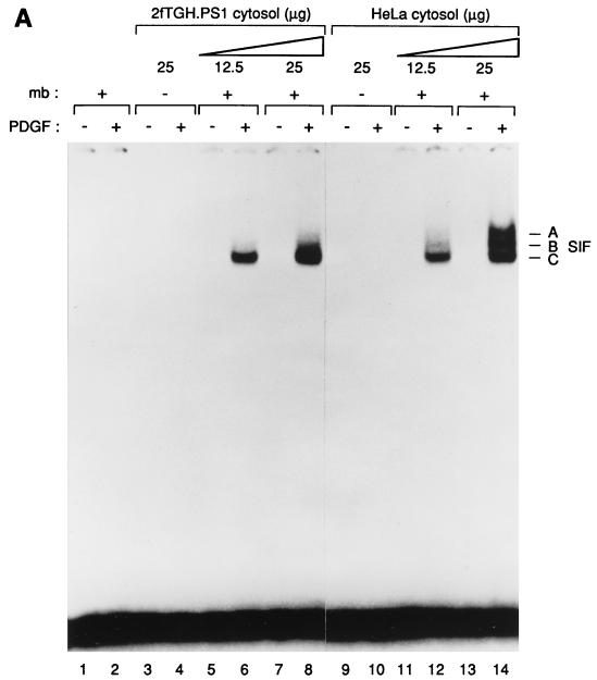
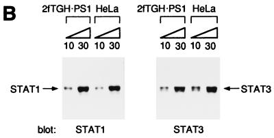
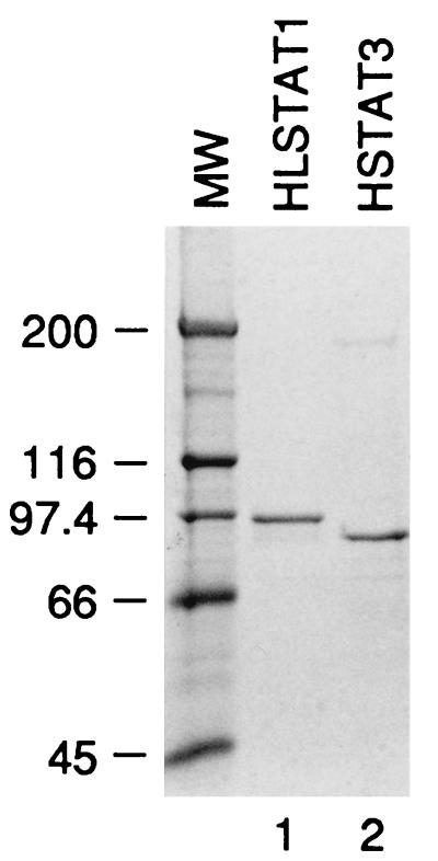
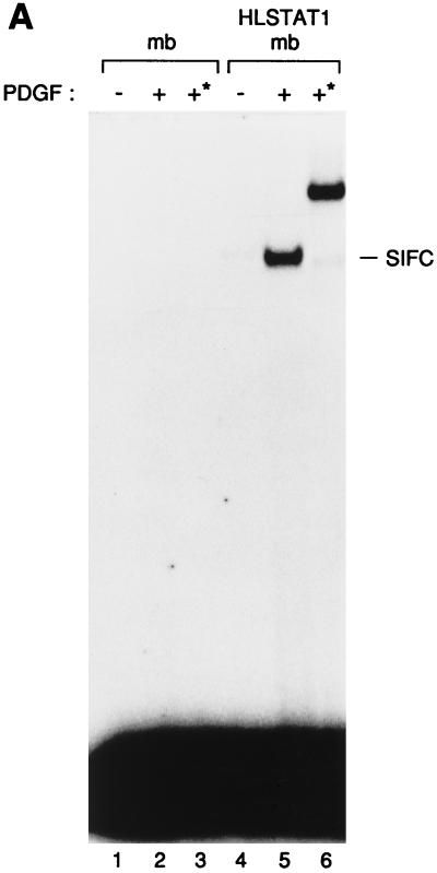
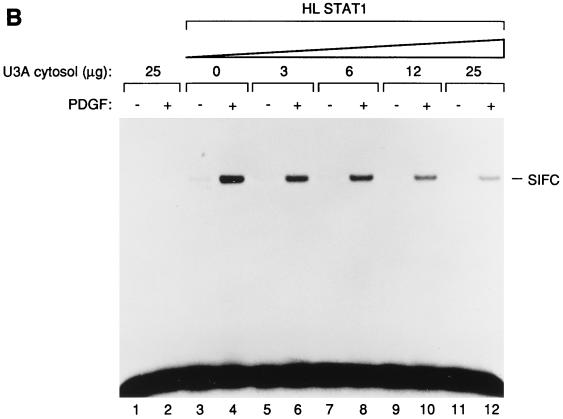
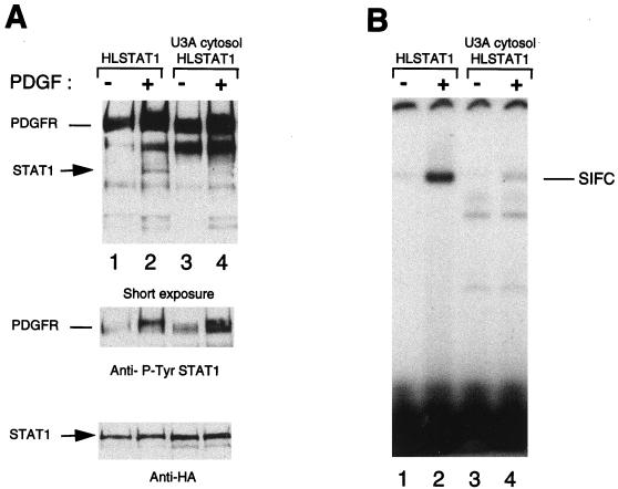
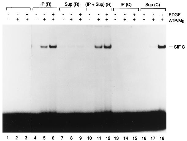
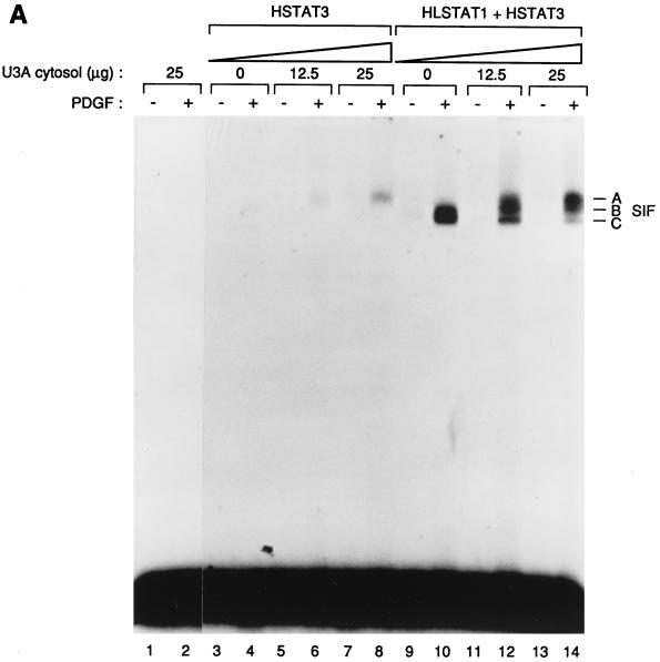
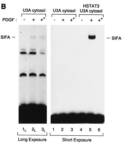
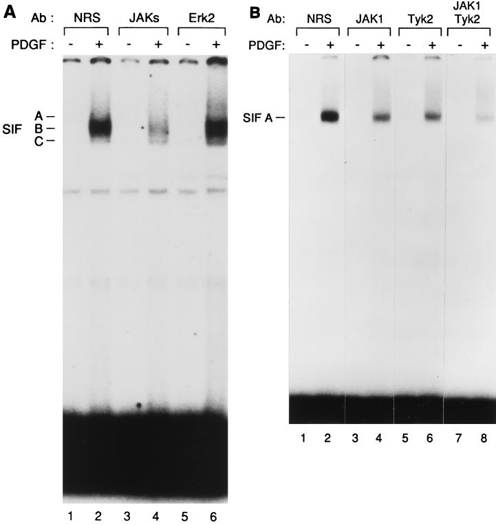
Ligand-dependent activation of the platelet-derived growth factor receptor (PDGFR) in fibroblasts in culture leads to the activation of the JAK family of protein-tyrosine kinases and of the transcription factors Stat1 and Stat3. To determine the biochemical mechanism of STAT activation by PDGFR, we devised a cell-free system composed of a membrane fraction from cells overexpressing PDGFR. When supplemented with crude cytosol, the membrane fraction supported PDGF- and ATP-dependent activation of both Stat1 and Stat3. However, the extent of Stat3 activation differed depending on the source of the cytosolic fraction. Using purified recombinant STAT proteins produced in Escherichia coli, we found that Stat1 could be activated by immunopurified PDGFR and showed no additional requirement for membrane- or cytosol-derived proteins. In contrast, activation of Stat3 exhibited a strong requirement for the cytosolic fraction. The activity present in the cytosolic fraction could be depleted with antibodies to JAK proteins. We conclude that the mechanisms of activation of Stat1 and Stat3 by PDGFR are distinct. Stat1 activation appears to result from a direct interaction with the receptor, whereas Stat3 activation additionally requires JAK proteins.


Figures

FIG. 1
FIG. 1
(A) PDGF-dependent activation of Stat1 and Stat3 in vitro. Detergent-solubilized membrane proteins from 2fTGH.PS1 cells, which overexpress the PDGF-β receptor, were mixed with cytosolic extracts from 2fTGH.PS1 or HeLa cells. The proteins were subjected to an in vitro kinase reaction in the presence of ATP and magnesium, and the activated STATs were analyzed by mobility shift assay using the high-affinity SIEm67 32P-labeled probe. Lanes: 1 and 2, no cytosolic extract; 3 to 8, 2fTGH.PS1 cytosol; 9 to 14, HeLa cytosol. The kinase reactions were done with (+) or without (−) membranes (mb) as indicated and in the presence (+) or absence (−) of PDGF as indicated. The three specific complexes SIF-A, SIF-B, and SIF-C are indicated at right. (B) 2fTGH.PS1 and HeLa cells express similar concentrations of Stat1 as well as of Stat3 proteins. Equal amounts (10 and 30 μg, respectively) of cytosolic extracts from 2fTGH.PS1 or HeLa cells were analyzed by Western blotting using antibodies directed against either Stat1 or Stat3. The STAT protein recognized by the antibodies is indicated.
FIG. 1
FIG. 1
(A) PDGF-dependent activation of Stat1 and Stat3 in vitro. Detergent-solubilized membrane proteins from 2fTGH.PS1 cells, which overexpress the PDGF-β receptor, were mixed with cytosolic extracts from 2fTGH.PS1 or HeLa cells. The proteins were subjected to an in vitro kinase reaction in the presence of ATP and magnesium, and the activated STATs were analyzed by mobility shift assay using the high-affinity SIEm67 32P-labeled probe. Lanes: 1 and 2, no cytosolic extract; 3 to 8, 2fTGH.PS1 cytosol; 9 to 14, HeLa cytosol. The kinase reactions were done with (+) or without (−) membranes (mb) as indicated and in the presence (+) or absence (−) of PDGF as indicated. The three specific complexes SIF-A, SIF-B, and SIF-C are indicated at right. (B) 2fTGH.PS1 and HeLa cells express similar concentrations of Stat1 as well as of Stat3 proteins. Equal amounts (10 and 30 μg, respectively) of cytosolic extracts from 2fTGH.PS1 or HeLa cells were analyzed by Western blotting using antibodies directed against either Stat1 or Stat3. The STAT protein recognized by the antibodies is indicated.
FIG. 2
FIG. 2
Purification of Stat1 and Stat3. Histidine-tagged Stat1 and Stat3 proteins were expressed in E. coli and purified on nickel. The purified proteins were analyzed by polyacrylamide gel electrophoresis and revealed by Coomassie staining. Stat3 harbors the histidine tag at its N terminus (HSTAT3) while Stat1 contains an additional hemagglutinin tag between the polyhistidine sequence and the N terminus of the protein (HLSTAT1). The molecular weight (MW) markers are indicated (in thousands) on the left of the gel.
FIG. 3
FIG. 3
(A) In vitro activation of the purified Stat protein. The purified Stat1 protein was mixed with 2fTGH.PS1 membranes (mb), activated in vitro, and tested by EMSA on an SIE probe (lanes 4 to 6). Lanes 1 to 3, mock reactions with membranes only. The kinase reactions were done in the presence (+) or absence (−) of PDGF. The addition of the 12CA5 monoclonal antibody during the gel shift reaction is indicated by an asterisk. The SIF-C complex corresponding to the binding of Stat1 to the SIE site is indicated at the right. (B) The PDGF-dependent activation of Stat1 is inhibited by cytosolic proteins. The purified HLSTAT1 protein was mixed with increasing concentrations of U3A cytosolic extracts and subjected to the in vitro kinase reaction using 2fTGH.PS1 membrane fractions as the source of PDGF-β receptor. As a control, U3A cytosolic proteins (25 μg) were tested in the kinase reaction in the absence of recombinant Stat1 protein (lanes 1 and 2). The kinase reactions were done in the absence (−) or presence (+) of PDGF. The SIF-C complex is indicated.
FIG. 3
FIG. 3
(A) In vitro activation of the purified Stat protein. The purified Stat1 protein was mixed with 2fTGH.PS1 membranes (mb), activated in vitro, and tested by EMSA on an SIE probe (lanes 4 to 6). Lanes 1 to 3, mock reactions with membranes only. The kinase reactions were done in the presence (+) or absence (−) of PDGF. The addition of the 12CA5 monoclonal antibody during the gel shift reaction is indicated by an asterisk. The SIF-C complex corresponding to the binding of Stat1 to the SIE site is indicated at the right. (B) The PDGF-dependent activation of Stat1 is inhibited by cytosolic proteins. The purified HLSTAT1 protein was mixed with increasing concentrations of U3A cytosolic extracts and subjected to the in vitro kinase reaction using 2fTGH.PS1 membrane fractions as the source of PDGF-β receptor. As a control, U3A cytosolic proteins (25 μg) were tested in the kinase reaction in the absence of recombinant Stat1 protein (lanes 1 and 2). The kinase reactions were done in the absence (−) or presence (+) of PDGF. The SIF-C complex is indicated.
FIG. 4
FIG. 4
Stat1 tyrosine phosphorylation and DNA binding activity are both inhibited by cytosolic proteins. The purified HLSTAT1 protein was subjected to the in vitro kinase reaction using 2fTGH.PS1 membranes (40 μg), in the absence (−) or presence (+) of U3A cytosolic extract (100 μg). Following the kinase reaction, half of the reaction mixture was analyzed by Western blotting using Stat1 phosphotyrosine (Y701) antibodies (A), the second half of the reaction mixture was analyzed by EMSA on an SIE probe (B). The phosphorylated Stat1 protein is indicated. Reprobing of the blot with an antihemagglutinin antibody (12CA5) shows equal Stat1 concentrations in the various lanes.
FIG. 5
FIG. 5
Activation of Stat1 protein by the immunoprecipitated PDGF-β receptor. The PDGF-β receptor was immunoprecipitated from detergent-solubilized membranes of 2fTGH.PS1 cells. The purified Stat1 was incubated in the in vitro kinase reaction with the immunoprecipitated PDGFR [IP (R); lanes 4 to 6], the membrane fraction depleted of PDGFR [Sup (R); lanes 7 to 9] or a mixture of the two [(IP + Sup) (R); lanes 10 to 12] and tested by EMSA on an SIE probe. In the mock immunoprecipitations [IP (C); lanes 13 to 15] or depletions [Sup (C); lanes 16 to 18], normal rabbit serum was used instead of specific anti-PDGFR antibodies. Lanes 1 to 3, control reactions where Stat1 was incubated without membrane proteins. The kinase reactions were done in the presence (+) or absence (−) of PDGF as well as of ATP/magnesium. The SIF-C complex corresponding to Stat1 binding to the SIE site is indicated.
FIG. 6
FIG. 6
(A) In vitro activation of Stat3 by the PDGF receptor requires cytosolic proteins. The purified Stat3 protein (HSTAT3; lanes 3 to 8) or a mixture of Stat1 and Stat3 (HLSTAT1 + HSTAT3; lanes 9 to 14) were incubated with increasing concentrations of U3A cytosol, activated in vitro, and tested by EMSA on an SIE probe. As a control, the kinase reaction was done with U3A cytosol (25 μg), in the absence of recombinant STATs (lanes 1 and 2). Kinase reactions were done in the presence (+) or absence (−) of PDGF. The three specific complexes SIF-A, SIF-B, and SIF-C are indicated. (B) The SIF-A complex generated in vitro corresponds to the activation of the recombinant Stat3 protein. HSTAT3 was incubated with U3A cytosol for the kinase assay and tested by EMSA (lanes 4 to 6). Control reactions were performed in the absence of recombinant Stat3 (lanes 1 to 3). In lanes 3 and 6, protein extracts obtained by the kinase reactions were depleted on nickel resin of histidine-containing proteins prior to the EMSA. Depletion on nickel resin is indicated by an asterisk. The SIF-A complex obtained in the presence (+) of PDGF is indicated. The left panel shows a longer exposure of the gel (lanes 1L to 3L).
FIG. 6
FIG. 6
(A) In vitro activation of Stat3 by the PDGF receptor requires cytosolic proteins. The purified Stat3 protein (HSTAT3; lanes 3 to 8) or a mixture of Stat1 and Stat3 (HLSTAT1 + HSTAT3; lanes 9 to 14) were incubated with increasing concentrations of U3A cytosol, activated in vitro, and tested by EMSA on an SIE probe. As a control, the kinase reaction was done with U3A cytosol (25 μg), in the absence of recombinant STATs (lanes 1 and 2). Kinase reactions were done in the presence (+) or absence (−) of PDGF. The three specific complexes SIF-A, SIF-B, and SIF-C are indicated. (B) The SIF-A complex generated in vitro corresponds to the activation of the recombinant Stat3 protein. HSTAT3 was incubated with U3A cytosol for the kinase assay and tested by EMSA (lanes 4 to 6). Control reactions were performed in the absence of recombinant Stat3 (lanes 1 to 3). In lanes 3 and 6, protein extracts obtained by the kinase reactions were depleted on nickel resin of histidine-containing proteins prior to the EMSA. Depletion on nickel resin is indicated by an asterisk. The SIF-A complex obtained in the presence (+) of PDGF is indicated. The left panel shows a longer exposure of the gel (lanes 1L to 3L).
FIG. 7
FIG. 7
(A) JAKs are required for Stat3 activation. A mixture of purified Stat1 and Stat3 recombinant proteins was incubated and activated in vitro with U3A cytosolic extracts that had been previously incubated with a cocktail of antibodies (Ab) to JAKs (lanes 3 and 4), with antibodies to ERK2 (lanes 5 and 6), or, as a control, with normal rabbit serum (NRS) (lanes 1 and 2). The activated STATs were then tested by EMSA on an SIE probe. The kinase reactions were done in the presence (+) or absence (−) of PDGF. The three specific complexes SIF-A, SIF-B, and SIF-C are indicated. (B) Individual JAKs contribute to Stat3 activation by the PDGF-β receptor. The purified Stat3 protein was incubated with U3A cytosol that had been previously depleted using antibodies to JAK1 (lanes 3 and 4), Tyk2 (lanes 5 and 6), a mixture of these two antibodies (lanes 7 and 8), or, as a control, with normal rabbit serum (lanes 1 and 2). The kinase reactions were done in the presence (+) or absence (−) of PDGF. The SIF-A complex obtained by EMSA on an SIE probe is indicated.

References

    1. Aman M J, Leonard W J. Cytokine signaling: cytokine-inducible signaling inhibitors. Curr Biol. 1997;7:784–788. - PubMed
    1. Bach E A, Auguet M, Schreiber R D. The IFNγ receptor: a paradigm for cytokine receptor signaling. Annu Rev Immunol. 1997;15:563–591. - PubMed
    1. Briscoe J, Kohlhuber F, Müller M. JAKs and STATs branch out. Trends Cell Biol. 1996;6:336–340. - PubMed
    1. Darnell J E., Jr STATs and gene regulation. Science. 1997;277:1630–1635. - PubMed
    1. Darnell J J, Kerr I M, Stark G R. Jak-STAT pathways and transcriptional activation in response to IFNs and other extracellular signaling proteins. Science. 1994;264:1415–1421. - PubMed

Publication types

MeSH terms