. 1999 May;19(5):3769-78.
doi: 10.1128/MCB.19.5.3769.

TAFII40 protein is encoded by the e(y)1 gene: biological consequences of mutations

Affiliations

TAFII40 protein is encoded by the e(y)1 gene: biological consequences of mutations

A Soldatov et al. Mol Cell Biol. 1999 May.

Abstract

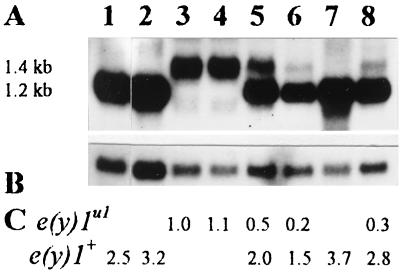
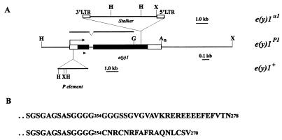
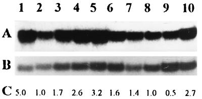
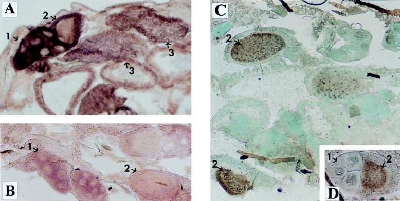
The enhancer of yellow 1 gene, e(y)1, of Drosophila melanogaster has been cloned and demonstrated to encode the TAFII40 protein. The e(y)1 gene is expressed in females much more strongly than in males due to the accumulation of e(y)1 mRNA in the ovaries. Two different e(y)1 mutations have been obtained. The e(y)1(ul) mutation, induced by the insertion of Stalker into the coding region, leads to the replacement of 25 carboxy-terminal amino acids by 17 amino acids encoded by the Stalker sequences and to a decrease of the e(y)1 transcription level. The latter is the main cause of dramatic underdevelopment of the ovaries and sterility of females bearing the e(y)1 mutation. This follows from the restoration of female fertility upon transformation of e(y)1(u1) flies with a construction synthesizing the mutant protein. The e(y)1(P1) mutation induced by P element insertion into the transcribed nontranslated region of the gene has almost no influence on the phenotype of flies. However, in combination with the phP1 mutation, which leads to a strong P element-mediated suppression of e(y)1 transcription, this mutation is lethal. Genetic studies of the e(y)1(u1) mutation revealed a sensitivity of the yellow and white expression to the TAFII40/e(y)1 level. The su(Hw)-binding region, Drosophila insulator, stabilizes the expression of the white gene and makes it independent of the e(y)1(u1) mutation.


Figures

FIG. 1
FIG. 1
Transcription of the e(y)1u1 and e(y)1+ genes. (A) Northern blot hybridization of the fragment of e(y)1/TAFII40 cDNA (Fig. 2A) with mRNA from Oregon R males (lane 1), Oregon R embryos (lane 2), e(y)1u1/Y males (lane 3), e(y)1u1/e(y)1u1 females (lane 4), e(y)1u1/e(y)1+ females (lane 5), embryos from the ♀ e(y)1u1/e(y)1+ × ♂ e(y)1u1/Y cross (lane 6), C(1)RM,1yf females (lane 7), and embryos from the ♀ C(1)RM,yf × ♂ e(y)1u1/Y cross (lane 8). (B) The same blot hybridized with the Ras2 probe. (C) Relative level of e(y)1/TAFII40 transcription. The Northern blot was analyzed on a PhosphoImager; signals were normalized according to the results of Ras2 hybridization. The level of transcription in e(y)1u1/Y males was taken as 1.
FIG. 2
FIG. 2
Structure of the e(y)1/TAFII40 gene. (A) Map of the e(y)1u1 mutation. Black boxes, the coding regions of e(y)1; open boxes, transcribed, nontranslated regions. The arrow indicates the direction of transcription. H, HindIII; X, XhoI; G, BglII. The region shown was used for wild-type phenotype rescue. The upper line indicates the region from the cDNA clone, which was used as a probe in Northern blot hybridization. The position of primers for RACE is indicated by a triangle. (B) Amino acid sequence of the carboxy terminus of wild-type (upper line) and mutant (lower line) TAFII40 protein.
FIG. 3
FIG. 3
Transcription of the e(y)1/TAFII40 gene at different stages of development of D. melanogaster. (A) Northern blot hybridization of a fragment of e(y)1/TAFII40 cDNA (Fig. 2A) with mRNA from the Oregon R strain. Samples are from adult females (lane 1) and males (lane 2); late (lane 3), middle (lane 4), and early (lane 5) pupae; late third (lane 6)-, early third (lane 7)-, second (lane 8)-, and first (lane 9)-instar larvae; and embryos (lane 10). (B) The same blot, hybridized with the Ras2 probe. (C) Relative level of e(y)1/TAFII40 transcription. Signals were normalized according to the results of Ras2 hybridization. The level of e(y)1 transcription in males was taken as 1.
FIG. 4
FIG. 4
Expression of e(y)1 in different tissues of Oregon R flies. (A and B) In situ hybridization of a frontal tissue section of female abdomen with the DIG-labeled e(y)1 antisense (A) and sense (B) RNA probes. (C and D) Immunostaining of a frontal tissue section of female abdomen with antibodies to e(y)1 protein. Horseradish peroxidase and DAB were used for visualization; the tissue was counterstained with fast green. One can see a high level of e(y)1 transcription and expression in ovaries: 1, in trophocytes; 2, in primary oocytes; 3, in mature oocytes. Note that while the level of e(y)1 mRNA content is high in trophocytes and mature oocytes and low in primary oocytes, the TAFII40 protein is predominantly detected in oocytes rather than in trophocytes (C). Magnification, ×130.
FIG. 5
FIG. 5
Western blot analysis of e(y)1 expression. Shown are results of immunoprecipitation of e(y)1 and e(y)1u1 proteins from nuclear extracts from adult flies of the Oregon R (lane 1), e(y)1u1 (lanes 2 to 4), and e(y)1+ strains (lanes 5 to 7) and the recombinant His-tagged protein (lane 8). The positions of e(y)1 and e(y)1u1 proteins are shown on the left.
FIG. 6
FIG. 6
Immunostaining of polytene chromosomes from wild-type Oregon R (A) and e(y)1u1 (B) larvae with antibodies against e(y)1 and Cy3-conjugated secondary antibodies. Original magnification, ×1,000.
FIG. 7
FIG. 7
Effect of the e(y)1u1 mutation on e(y)1 transcription. (A) Northern blot hybridization of the fragment of e(y)1/TAFII40 cDNA (Fig. 2A) with mRNA isolated at different stages of development of the progeny of the ♀ C(1)RM, yf × ♂ e(y)1u1/Y cross: males (lane 1); late (lane 2), middle (lane 3), and early (lane 4) pupae; third (lane 5)- and first (lane 6)-instar larvae; embryos (lane 7); and adult females (lane 8). Lane 9, mRNA from females of the Oregon R strain. (B) The same blot, hybridized with the Ras2 probe.
FIG. 8
FIG. 8
Ovaries from wild-type (A) and e(y)1u1 (B) flies. Immunostaining of frontal tissue section of female abdomen with antibodies to the e(y)1 protein. Horseradish peroxidase and Sigma fast DAB with a metal enhancer were used for visualization. Magnification, ×70.
FIG. 9
FIG. 9
Ovaries from wild-type (A) and e(y)1u1 (B and C) flies (total preparation). Magnification, ×40.
FIG. 10
FIG. 10
Genetic analysis of interaction of y alleles with e(y)1u1 mutation. The schemes for y alleles are not to scale. yellow transcripts are shown by arrows; transcriptional enhancers are indicated by shaded ovals. The enhancers that control yellow expression in the wings and body cuticle are located in the 5′-upstream region of the yellow gene, whereas enhancers controlling yellow expression in the bristles reside in the intron of the gene (22). The su(Hw)-binding region is indicated by empty boxes; insertions found in the various alleles are represented by triangles. The total number of circles in the phenotype column indicates the levels of pigmentation of the body and wings (column 1), thoracic bristles (column 2), leg bristles (column 3), and abdominal bristles (column 4). The number of black circles shows the inhibitory effect of the e(y)1u1 mutation on yellow expression for different y alleles. Each circle represents one point on the scale described in the footnote to Table 1.

Similar articles

Cited by

References

    1. Altschul S F, Gish W, Miller W, Myers E W, Lipman D J. Basic local alignment search tool. J Mol Biol. 1990;215:403–410. - PubMed
    1. Apone L M, Virbasius C-M A, Reese J C, Green M R. Yeast TAFII90 is required for cell-cycle progression through G2/M but not for general transcription activation. Genes Dev. 1996;10:2368–2380. - PubMed
    1. Ashburner M. Drosophila: a laboratory manual. Cold Spring Harbor, N.Y: Cold Spring Harbor Laboratory; 1989.
    1. Belenkaya T, Barseguyan K, Hovhannisayn H, Biryukova I, Kochieva E Z, Georgiev P. P element sequences can compensate a deletion of the yellow regulatory region in Drosophila melanogaster. Mol Gen Genet. 1998;259:79–87. - PubMed
    1. Belenkaya T, Soldatov A, Nabirochkina E, Birjukova I, Georgieva S, Georgiev P. The allele of the polyhomeotic gene induced by P element insertion encodes a new chimeric protein, that negatively regulates the expression of P-induced alleles in the yellow locus of Drosophila melanogaster. Genetics. 1998;150:687–697. - PMC - PubMed

Publication types

LinkOut - more resources