. 1999 May;19(5):3779-87.
doi: 10.1128/MCB.19.5.3779.

The Saccharomyces cerevisiae homologues of endonuclease III from Escherichia coli, Ntg1 and Ntg2, are both required for efficient repair of spontaneous and induced oxidative DNA damage in yeast

Affiliations

The Saccharomyces cerevisiae homologues of endonuclease III from Escherichia coli, Ntg1 and Ntg2, are both required for efficient repair of spontaneous and induced oxidative DNA damage in yeast

I Alseth et al. Mol Cell Biol. 1999 May.

Abstract

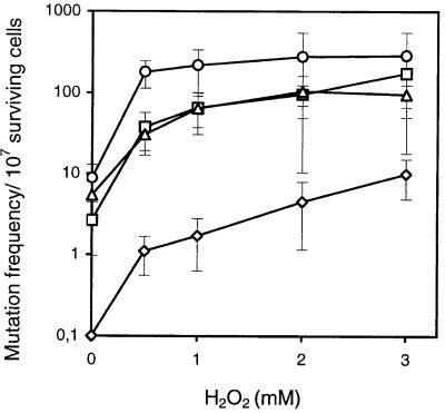
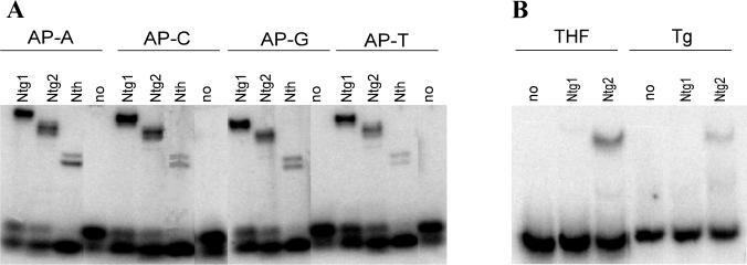
Endonuclease III from Escherichia coli is the prototype of a ubiquitous DNA repair enzyme essential for the removal of oxidized pyrimidine base damage. The yeast genome project has revealed the presence of two genes in Saccharomyces cerevisiae, NTG1 and NTG2, encoding proteins with similarity to endonuclease III. Both contain the highly conserved helix-hairpin-helix motif, whereas only one (Ntg2) harbors the characteristic iron-sulfur cluster of the endonuclease III family. We have characterized these gene functions by mutant and enzyme analysis as well as by gene expression and intracellular localization studies. Targeted gene disruption of NTG1 and NTG2 produced mutants with greatly increased spontaneous and hydrogen peroxide-induced mutation frequency relative to the wild type, and the mutation response was further increased in the double mutant. Both enzymes were found to remove thymine glycol and 2, 6-diamino-4-hydroxy-5-N-methylformamidopyrimidine (faPy) residues from DNA with high efficiency. However, on UV-irradiated DNA, saturating concentrations of Ntg2 removed only half of the cytosine photoproducts released by Ntg1. Conversely, 5-hydroxycytosine was removed efficiently only by Ntg2. The enzymes appear to have different reaction modes, as judged from much higher affinity of Ntg2 for damaged DNA and more efficient borhydride trapping of Ntg1 to abasic sites in DNA despite limited DNA binding. Northern blot and promoter fusion analysis showed that NTG1 is inducible by cell exposure to DNA-damaging agents, whereas NTG2 is constitutively expressed. Ntg2 appears to be a nuclear enzyme, whereas Ntg1 was sorted both to the nucleus and to the mitochondria. We conclude that functions of both NTG1 and NTG2 are important for removal of oxidative DNA damage in yeast.


Figures

FIG. 1
FIG. 1
Alignment of the S. cerevisiae Ntg1 and Ntg2 proteins. Highlighted amino acids represent identical residues. Different structural features are underlined above and below the alignment as indicated with reference to Ntg1 and Ntg2, respectively. NLS, nuclear localization signal; MLS, mitochondrial localization signal.
FIG. 2
FIG. 2
Spontaneous and H2O2-induced mutation frequencies of S. cerevisiae mutants lacking Ntg1 and/or Ntg2. The frequency of mutations to canavanine resistance in S. cerevisiae FF18733 (wild type; ◊), RH101 (ntg1::URA3; □), RH102 (ntg2::LEU2; ▵), and RH103 (ntg1::URA3, ntg2::LEU2; ○) was measured in nontreated cells and cells exposed to H2O2 at the doses indicated. The level of spontaenous mutations plotted for wild-type cells is an upper estimate of the mutation frequency, as no mutants were detected when 108 cells were plated on the canavanine plates.
FIG. 3
FIG. 3
SDS-PAGE of different protein fractions obtained during purification of Ntg1 and Ntg2. (A) Ntg1. Samples include molecular weight standards (lanes 1 and 7), MonoQ fraction (lane 2), DNA-cellulose fraction (lane 3), MonoS fraction (lane 4), Affi-Gel Blue (AGB) fraction (lane 5), and cell extract (lane 6). (B) Ntg2. Samples include molecular weight standards (lanes 1 and 8), Superdex-75 fraction (lane 2), MonoS fraction (lane 3), DNA-cellulose fraction (lane 4), MonoQ fraction (lane 5), Affi-Gel Blue fraction (lane 6), and cell extract (lane 7).
FIG. 4
FIG. 4
Release of faPy residues by Ntg1 and Ntg2. Excision of faPy from [3H]faPy-poly(dG-dC)DNA (0.4 μg) was measured after enzyme incubation for 30 min at 37°C with increasing amounts of purified Ntg1(□), Ntg2 (▵), Fpg (◊), and Nth (○).
FIG. 5
FIG. 5
Activities of Ntg1 and Ntg2 on UV-irradiated DNA. (A) Enzyme cleavage of a 33P-end-labeled UV-irradiated DNA fragment (250 fmol) incubated with an excess amount of Ntg1 and Ntg2 for 30 min at 37°C. The cleavage products were separated on a DNA sequencing gel alongside DNA sequencing reactions. The type of base acted upon for each band position was identified as indicated, and band intensities were quantified by phosphorimaging (Ntg1 [open bars] and Ntg2 [filled bars]). (B) Release of radiolabeled cytosines from UV-irradiated [3H]C-DNA by increasing amounts of purified Ntg1 (□), Ntg2 (▵), and Nth (○) after incubation for 30 min at 37°C.
FIG. 6
FIG. 6
Cleavage of Tg- and 5-OHC-containing DNA. (A) Duplex 37-mer oligodeoxyribonucleotide containing a single Tg at position 19 (10 fmol) was incubated for 30 min at 37°C with 1 and 5 ng of Nth (lanes 1 and 2), Ntg1 (lanes 3 and 4), Ntg2 (lanes 5 and 6), or Nfo (lanes 7 and 8) or no enzyme (lane 9). Cleaved DNA was separated from intact DNA by 20% denaturing PAGE and visualized by phosphorimaging. (B) A duplex 26-mer containing 5-OHC at position 13 (10 fmol) was incubated for 30 min at 37°C with 5 and 20 ng each of Nth, Ntg1, Ntg2, or Nfo or no enzyme as indicated. Cleavage products were analyzed by 20% denaturing PAGE and phosphoimaging. nt, nucleotides.
FIG. 7
FIG. 7
Borhydride trapping and DNA binding analysis of Ntg1 and Ntg2. (A) Probing for covalent enzyme–AP-DNA intermediates by NaCNBH3 reduction. Duplex 32P-labeled oligodeoxyribonucleotide (80 fmol) containing a single AP site opposite A (lanes 1 to 4), C (lanes 5 to 8), G (lanes 9 to 12), and T (lanes (13 to 16) was incubated with 40 ng of Ntg1 (lanes 1, 5, 9, and 13), Ntg2 (lanes 2, 6, 10, and 14), or Nth (lanes 3, 7, 11, and 15) or without enzyme (lane 4, 8, 12, and 16). Protein-DNA complexes were separated from DNA by 10% Tricine-SDS-PAGE. (B) Duplex DNA containing a single THF (10 fmol; lanes 1 to 3) or Tg (50 fmol; lanes 4 to 6) residue was incubated with 5 ng of Ntg1 (lanes 2 and 5), 5 ng of Ntg2 (lanes 3 and 6), or no enzyme (lanes 1 and 4) for 15 min at 4°C and analyzed by 10% nondenaturing PAGE.
FIG. 8
FIG. 8
Fluorescence microscopy of S. cerevisiae expressing fusions of Ntg1- and Ntg2 to GFP. S. cerevisiae FF18733 was transformed by DNA constructs expressing GFP alone, NTG1-GFPn, NTG1-GFPc, or NTG2-GFPc. Cells were also stained with DAPI to visualize the DNA in the nucleus and the mitochondria.
FIG. 9
FIG. 9
Expression of NTG1 and NTG2 in S. cerevisiae exposed to DNA-damaging agents. (A) Northern blot analysis. Total RNA isolated from untreated cells and cells exposed to H2O2 (1 mM), menadione (5 mM), MMS (0.05%) and NQO (2 μg/ml) was size separated on a formaldehyde–1% agarose gel and blotted onto a nylon membrane. The blot was hybridized with a 1.2-kb NTG1 probe, stripped, rehybridized with a 1.1-kb NTG2 probe, stripped again, and finally hybridized with a 2.1-kb β-actin probe. Quantification and calculation of the hybridization signals of NTG1/NTG2 relative to β-actin (normalized to 1 for the nontreated samples) were 1.2/0.8, 2.3/0.7, 1.9/1.2, and 1.9/1.3 for H2O2, menadione, MMS, and NQO, respectively. (B) Promoter fusion analysis. Yeast FF18733 carrying the NTG1::lacZ or the NTG2::lacZ fusion on a centromeric plasmid was assayed for β-galactosidase activity expressed in nontreated cells (open bars) or cells exposed to menadione (0.3 mM; grey bars) or MMS (0.05%; filled bars).

References

    1. Ames B N, Gold L S, Willett W C. The causes and prevention of cancer. Proc Natl Acad Sci USA. 1995;92:5258–5265. - PMC - PubMed
    1. Aspinwall R, Rothwell D G, Roldan-Arjona T, Anselmino C, Ward C J, Cheadle J P, Sampson J R, Lindahl T, Harris P C, Hickson I D. Cloning and characterization of a functional human homolog of Escherichia coli endonuclease III. Proc Natl Acad Sci USA. 1997;94:109–114. - PMC - PubMed
    1. Augeri L, Lee L, Barton A B, Doetsch P W. Purification, characterization, gene cloning, and expression of Saccharomyces cerevisiae redoxyendonuclease, a homolog of Escherichia coli endonuclease III. Biochemistry. 1997;36:721–729. - PubMed
    1. Barton A B, Kaback D B. Molecular cloning of chromosome I DNA from Saccharomyces cerevisiae: analysis of the genes in the FUN38-MAK16-SPO7 region. J Bacteriol. 1994;176:1872–1880. - PMC - PubMed
    1. Bjørås M, Luna L, Johnsen B, Hoff E, Haug T, Rognes T, Seeberg B. Opposite base-dependent reactions of a human base excision repair enzyme on DNA containing 7,8-dihydro-8-oxoguanine and abasic sites. EMBO J. 1997;16:6314–6322. - PMC - PubMed

Publication types

MeSH terms