. 1999 May;19(5):3808-15.
doi: 10.1128/MCB.19.5.3808.

HERF1, a novel hematopoiesis-specific RING finger protein, is required for terminal differentiation of erythroid cells

Affiliations

HERF1, a novel hematopoiesis-specific RING finger protein, is required for terminal differentiation of erythroid cells

H Harada et al. Mol Cell Biol. 1999 May.

Abstract

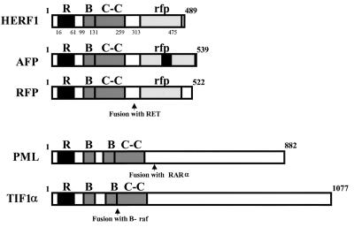
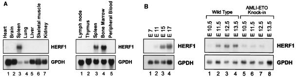
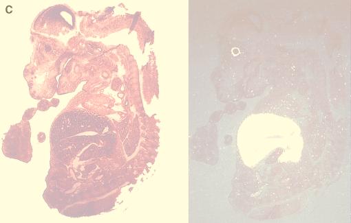
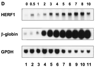
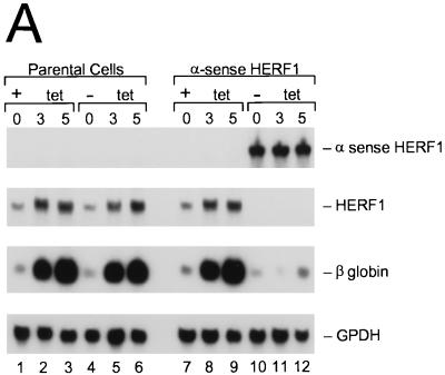
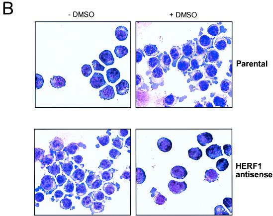
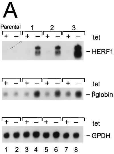
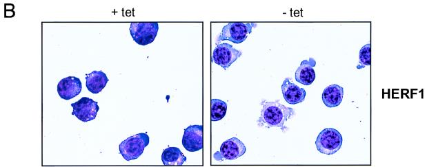
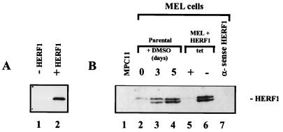
The AML1/core binding factor beta (CBFbeta) transcription factor is essential for definitive hematopoiesis; however, the downstream pathways through which it functions remain incompletely defined. Using a differential cloning approach to define components of this pathway, we have identified a novel gene designated HERF1 (for hematopoietic RING finger 1), whose expression during development is dependent on the presence of functional AML1/CBFbeta. HERF1 contains a tripartite RING finger-B box-alpha-helical coiled-coil domain and a C-terminal region homologous to the ret proto-oncogene-encoded finger protein. Expression of HERF1 during embryogenesis coincides with the appearance of definitive erythropoiesis and in adult mice is restricted to erythroid cells, increasing 30-fold during terminal differentiation. Importantly, inhibition of HERF1 expression blocked terminal erythroid differentiation of the murine erythroleukemia cell line MEL, whereas its overexpression induced erythroid maturation. These results suggest an important role for this protein in erythropoiesis.


Figures

FIG. 1
FIG. 1
Structural organization of selected members of the RING (R), B box (B), coiled-coil (C-C) family of proteins. RAR, retinoic acid receptor alpha.
FIG. 2
FIG. 2
Expression pattern of HERF1. Northern blots of adult tissues (A), murine embryos (B), or MEL cells following treatment with the differentiation-inducing agent DMSO (D) were hybridized with a full-length HERF1 cDNA. Filters were stripped and rehybridized with a probe for glycerol-3-phosphate dehydrogenase (GPDH) to assess the integrity and amount of RNA and with a probe for β-globin (D; numbers above the lanes represent days in DMSO). (C) In situ hybridization was performed with a 271-bp restriction fragment from the unique 3′ region of HERF1 labeled with [33P]UTP. The left side shows the bright-field view of a 12-μm sagittal section from an E15 embryo stained with hematoxylin and eosin; the right side is a dark-field view of the same section hybridized with the HERF1 probe. Sense control shows no specific hybridization (data not shown).
FIG. 2
FIG. 2
Expression pattern of HERF1. Northern blots of adult tissues (A), murine embryos (B), or MEL cells following treatment with the differentiation-inducing agent DMSO (D) were hybridized with a full-length HERF1 cDNA. Filters were stripped and rehybridized with a probe for glycerol-3-phosphate dehydrogenase (GPDH) to assess the integrity and amount of RNA and with a probe for β-globin (D; numbers above the lanes represent days in DMSO). (C) In situ hybridization was performed with a 271-bp restriction fragment from the unique 3′ region of HERF1 labeled with [33P]UTP. The left side shows the bright-field view of a 12-μm sagittal section from an E15 embryo stained with hematoxylin and eosin; the right side is a dark-field view of the same section hybridized with the HERF1 probe. Sense control shows no specific hybridization (data not shown).
FIG. 3
FIG. 3
Inhibition of DMSO-induced MEL cell differentiation by expression of α-sense HERF1. MEL cells expressing the tetracycline-regulated Tet-VP16 fusion protein, stably transfected with either an empty vector (parental) or a tetracycline-regulated α-sense HERF1-containing plasmid (α sense HERF1), were treated for 3 to 5 days with DMSO in the presence (+) or absence (−) of tetracycline (tet). Following the indicated treatments, cells were isolated and assessed by Northern blot analysis (A) and morphology (B). Northern blots of total RNA were sequentially hybridized with probes specific for the genes listed on the right side.
FIG. 3
FIG. 3
Inhibition of DMSO-induced MEL cell differentiation by expression of α-sense HERF1. MEL cells expressing the tetracycline-regulated Tet-VP16 fusion protein, stably transfected with either an empty vector (parental) or a tetracycline-regulated α-sense HERF1-containing plasmid (α sense HERF1), were treated for 3 to 5 days with DMSO in the presence (+) or absence (−) of tetracycline (tet). Following the indicated treatments, cells were isolated and assessed by Northern blot analysis (A) and morphology (B). Northern blots of total RNA were sequentially hybridized with probes specific for the genes listed on the right side.
FIG. 4
FIG. 4
Induction of MEL cell differentiation by enforced HERF1 expression. MEL cells expressing the tetracycline-regulated Tet-VP16 fusion protein, either with an empty plasmid vector (parental) or with a tetracycline-regulated HERF1-containing plasmid (clones 1 to 3), were grown in the presence (+) or absence (−) of tetracycline (tet). Following the indicated treatments, cells were isolated and analyzed by Northern blot analysis (A) and morphology (B). Northern blots were sequentially hybridized with probes specific for the genes listed on the right side.
FIG. 4
FIG. 4
Induction of MEL cell differentiation by enforced HERF1 expression. MEL cells expressing the tetracycline-regulated Tet-VP16 fusion protein, either with an empty plasmid vector (parental) or with a tetracycline-regulated HERF1-containing plasmid (clones 1 to 3), were grown in the presence (+) or absence (−) of tetracycline (tet). Following the indicated treatments, cells were isolated and analyzed by Northern blot analysis (A) and morphology (B). Northern blots were sequentially hybridized with probes specific for the genes listed on the right side.
FIG. 5
FIG. 5
Western blot analysis of HERF1, performed with an anti-HERF1 rabbit serum on total cell lysates prepared from Cos cells transfected with an empty vector (−HERF1) or a HERF1 expression plasmid (+HERF1) (A) or the B-lineage leukemic cell line MPC11, parental MEL cells treated with the differentiation-inducing agent DMSO, and MEL cells transfected with a tetracycline (tet)-regulated sense or α-sense HERF1 cDNA (B).

Similar articles

Cited by

References

    1. Bellini M, Lacroix J-C, Gall J G. A putative zinc-binding protein on lampbrush chromosome loops. EMBO J. 1993;12:107–114. - PMC - PubMed
    1. Borden K L B, Freemont P S. The RING finger domain: a recent example of a sequence-structure family. Curr Opin Struct Biol. 1996;6:395–401. - PubMed
    1. Borden K L B, Martin S R, O’Reilly N J, Lally J M, Reddy B A, Etkin L D, Freemont P S. Characterisation of a novel cysteine/histidine-rich metal binding domain from Xenopus nuclear factor XNF7. FEBS Lett. 1993;335:255–260. - PubMed
    1. Chang K S, Fan Y H, Andreeff M, Liu J, Mu Z M. The PML gene encodes a phosphoprotein associated with the nuclear matrix. Blood. 1995;85:3646–3653. - PubMed
    1. Chomczynski P, Sacchi N. Single-step method of RNA isolation by acid guanidinium thiocyanate-phenol-chloroform extraction. Anal Biochem. 1987;162:156–159. - PubMed

Publication types

MeSH terms

Associated data