. 1999 May;19(5):3895-903.
doi: 10.1128/MCB.19.5.3895.

Molecular determinants of the estrogen receptor-coactivator interface

Affiliations

Molecular determinants of the estrogen receptor-coactivator interface

H Y Mak et al. Mol Cell Biol. 1999 May.

Abstract

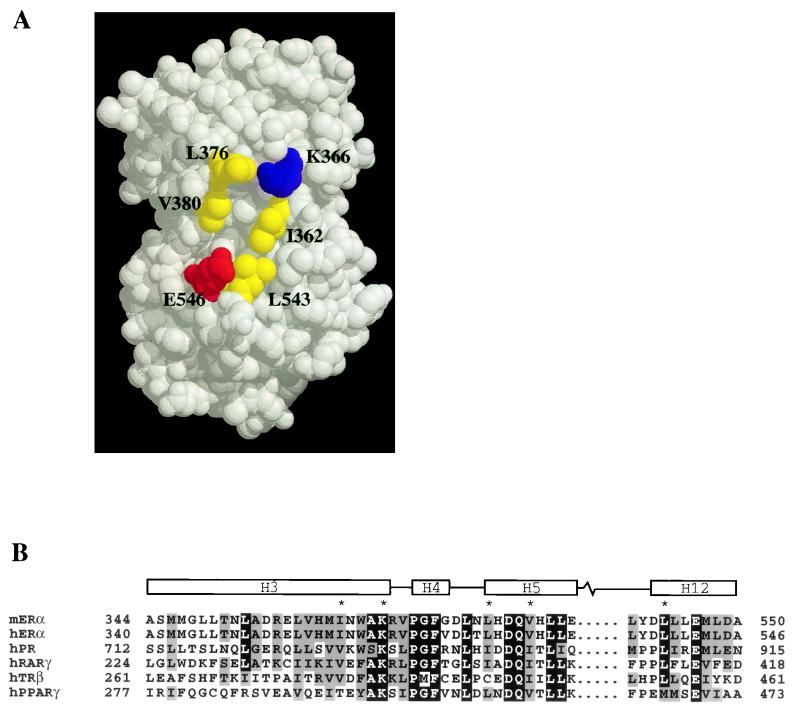
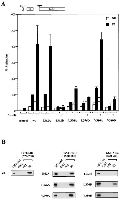
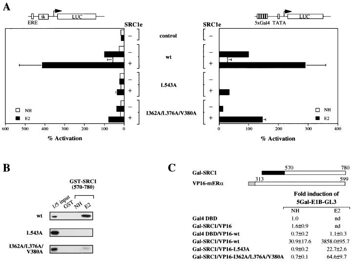
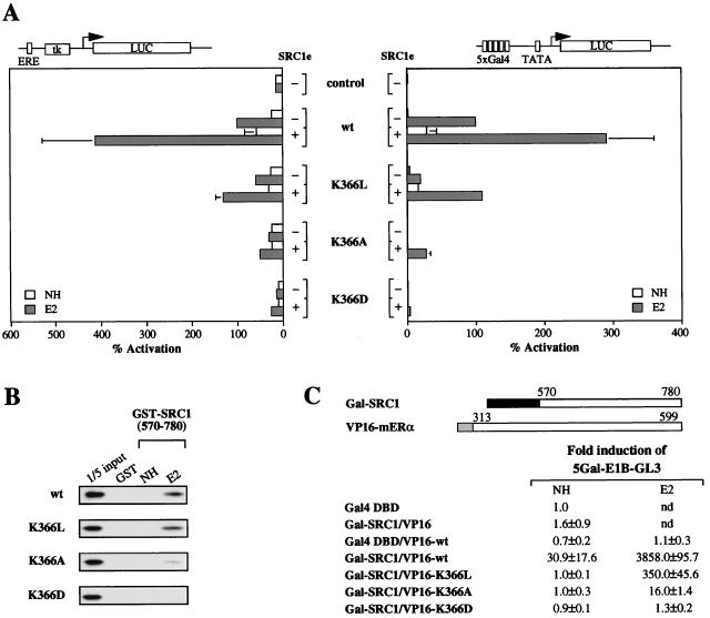
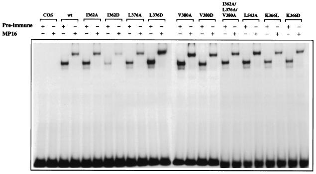
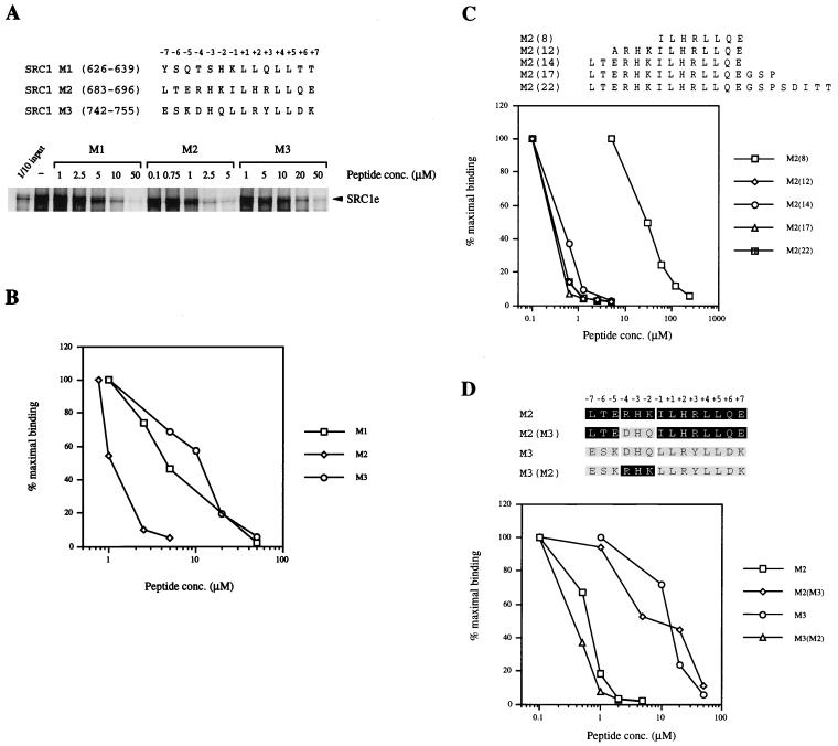
Transcriptional activation by the estrogen receptor is mediated through its interaction with coactivator proteins upon ligand binding. By systematic mutagenesis, we have identified a group of conserved hydrophobic residues in the ligand binding domain that are required for binding the p160 family of coactivators. Together with helix 12 and lysine 366 at the C-terminal end of helix 3, they form a hydrophobic groove that accommodates an LXXLL motif, which is essential for mediating coactivator binding to the receptor. Furthermore, we demonstrated that the high-affinity binding of motif 2, conserved in the p160 family, is due to the presence of three basic residues N terminal to the core LXXLL motif. The recruitment of p160 coactivators to the estrogen receptor is therefore likely to depend not only on the LXXLL motif making hydrophobic interactions with the docking surface on the receptor, but also on adjacent basic residues, which may be involved in the recognition of charged residues on the receptor to allow the initial docking of the motif.


Figures

FIG. 1
FIG. 1
Structure of the hERα LBD in the presence of 17β-estradiol. (A) Residues implicated in this study for participation in p160 coactivator binding are highlighted yellow (hydrophobic), red (acidic), and blue (basic). The residues are numbered as in mERα. The space-filled model was generated by RasMol and is based on the coordinates under the Protein Data Bank entry code 1ERE. (B) Sequence alignment of mERα LBD helices 3, 4, 5, and 12 with corresponding regions of members of the nuclear receptor superfamily whose agonist-bound crystal structures are solved. Note the absolute conservation of residues (marked with asterisks) in mERα and hERα, which are involved in coactivator binding. The boundaries for helices 3, 4, 5, and 12 are assigned according to the hERα LBD structure. The alignment was generated by Pileup (GCG) and formatted with MacBoxshade.
FIG. 2
FIG. 2
Surface hydrophobic residues in helices 3 and 5 of mERα are involved in coactivator binding. (A) COS-1 cells were transiently transfected with expression vector for wild-type (wt) or mutant receptors and pERE-tk-GL3 reporter in the absence (−) or presence (+) of 100 ng of full-length SRC1e. A cytomegalovirus promoter-driven pJ7-lacZ plasmid was cotransfected as the internal control. After transfection, cells were treated with ethanol vehicle alone (NH) or 17β-estradiol (E2) at 10−8 M for 24 h. Subsequently, cells were assayed for luciferase (LUC) and β-galactosidase activity. Normalized values are expressed as percentage of activity compared with that of wild-type mERα alone in the presence of β-estradiol (100%). The results shown represent the average of at least two independent experiments assayed in duplicate + standard errors. ERE, estrogen response element. (B) Binding activity of wild-type or mutant receptors to SRC1 in GST pull-down assay. In vitro-translated, [35S]methionine-labelled receptors were incubated with GST-SRC1 (aa 570 to 780) coupled to Sepharose beads in either the absence (NH) or presence (E2) of 10−6 M 17β-estradiol. Bound proteins were eluted and separated on SDS–10% polyacrylamide gels. Labelled proteins were detected by fluorography. The input lane represents 20% of the total volume of the lysate used in each reaction.
FIG. 3
FIG. 3
Functional analysis of L543A and I362A-L376A-V380A mutant receptors. (A) Wild-type (wt) or mutant full-length (left) or chimeric receptors consisting of the LBD of mERα fused to the DNA binding domain of Gal4 (right) were transiently transfected into COS-1 cells. Luciferase (LUC) reporter genes as indicated were cotransfected in the presence (+) or absence (−) of 100 ng of full-length SRC1e, and pJ7-lacZ was used as an internal control. Data are presented as described for Fig. 2A. ERE, estrogen response element. (B) Binding of mutant receptors to GST-SRC1 (aa 570 to 780) in vitro was examined under the same conditions as described for Fig. 2B. (C) In vivo interaction of mutant mERα LBDs with SRC1 (aa 570 to 780). The expression vectors used are schematically represented with the numbers indicating the amino acid position in the full-length protein. The darkly shaded box represents the Gal4 DNA binding domain (aa 1 to 147), and the lightly shaded box represents the activation domain of VP16 (aa 410 to 490). HeLa cells were transiently transfected with the indicated expression vectors, together with a p5Gal-E1B-GL3 reporter gene and the pJ7-lacZ internal control plasmid. Following transfection, cells were treated with ethanol vehicle alone (NH) or 10−8 M 17β-estradiol (E2). After 24 h, cell extracts were prepared and assayed for luciferase and β-galactosidase activities. Normalized values are expressed as fold induction compared with that of the Gal4 DNA binding domain alone (set as 1). The results shown represent the average of at least two independent experiments assayed in duplicate ± standard error. nd, not determined.
FIG. 4
FIG. 4
Mutation of K366 reveals its dual property in AF2 activity. (A) COS-1 cells were transiently transfected with expression vector for wild-type (wt) or mutant receptors (left) or Gal4-chimeric receptors (right), the pJ7-lacZ internal control plasmid, and the luciferase (LUC) reporter plasmid as indicated. Data are presented as described for Fig. 2A. (B) Binding of mutant receptors to GST-SRC1 (aa 570 to 780) was examined under the same conditions as described for Fig. 2B. (C) In vivo interaction of mutant mERα LBDs with SRC1 (aa 570 to 780) in transiently transfected HeLa cells. Data are presented as described for Fig. 3C. nd, not determined.
FIG. 5
FIG. 5
Mutant receptors bind to DNA with affinity similar to that of the wild-type receptor. Full-length wild-type (wt) or mutant receptors were transiently expressed in COS-1 cells. Equal amounts of receptor were analyzed for DNA binding in an electrophoretic mobility shift assay using a 32P-labelled oligonucleotide containing a single consensus estrogen response element from the vitellogenin A2 gene promoter. Binding reactions were performed either in the presence of ERα-specific antibody MP16 or preimmune serum. Protein-DNA complexes were separated on 6% native polyacrylamide gels and detected by autoradiography.
FIG. 6
FIG. 6
Differential inhibition of mERα-SRC1 interaction in vitro by an LXXLL motif containing peptides. (A) Comparison of peptides encompassing either SRC1 LXXLL motif 1, 2, or 3. A GST fusion protein of mERα LBD which had been coupled to Sepharose beads was incubated with in vitro-translated [35S]methionine-labelled SRC1e protein and increasing amounts of LXXLL motif-containing peptide, in the presence of 10−6 M 17β-estradiol. Bound labelled proteins were eluted, separated on SDS–10% polyacrylamide gels, and detected by fluorography. The input lane represents 10% of the total volume of the lysate used in each reaction. (B) Graphical representation of results from Fig. 6A. The amount of bound SRC1e protein was quantified with a PhosphorImager and is expressed as a percentage of maximal binding relative to the amount of bound proteins in the absence of any LXXLL motif-containing peptide (100%). At least two independent experiments were performed, and the data shown are from one representative experiment. (C) Effect of flanking residues on inhibition of mERα-SRC1 interaction by motif 2-containing peptide. Increasing amounts of M2 peptide, the length of which varied from 8 to 22 residues, were used to inhibit interaction between GST-mERα LBD and [35S]methionine-labelled SRC1e protein in an assay described for panel A. Data are presented as described for panel B and are from one representative experiment. At least two independent experiments were performed. (D) Three basic residues N terminal to LXXLL core motif 2 confer high-affinity binding to mERα. Residues −4DHQ−2 of SRC1 motif 3 were replaced by residues −4RHK−2 from the corresponding positions of motif 2 in a 14-mer peptide and vice versa. Increasing amounts of wild-type or mutant peptides were incubated with GST-mERα LBD and [35S]methionine-labelled SRC1e protein in an assay described for panel A. Data from one representative experiment are presented as described for panel B. At least two independent experiments were performed.

Similar articles

Cited by

References

    1. Anzick S L, Kononen J, Walker R L, Azorsa D O, Tanner M M, Guan X-Y, Sauter G, Kallioniemi O-P, Trent J M, Meltzer P S. AIB1, a steroid receptor coactivator amplified in breast and ovarian cancer. Science. 1997;277:965–968. - PubMed
    1. Beato M, Herrlich P, Schutz G. Steroid-hormone receptors—many actors in search of a plot. Cell. 1995;83:851–857. - PubMed
    1. Bourguet W, Ruff M, Chambon P, Gronemeyer H, Moras D. Crystal-structure of the ligand-binding domain of the human nuclear receptor RXR-alpha. Nature. 1995;375:377–382. - PubMed
    1. Brzozowski A M, Pike A C W, Dauter Z, Hubbard R E, Bonn T, Engstrom O, Ohman L, Greene G L, Gustafsson J-A, Carlquist M. Molecular basis of agonism and antagonism in the oestrogen receptor. Nature. 1997;389:753–758. - PubMed
    1. Butler A J, Parker M G. Coup-TF-II homodimers are formed in preference to heterodimers with RXR-alpha or TR-beta in intact-cells. Nucleic Acids Res. 1995;23:4143–4150. - PMC - PubMed

Publication types

MeSH terms