

A contiguous 3-Mb sequence-ready map in the S3-MX region on 21q22.2 based on high- throughput nonisotopic library screenings
- PMID: 10207158
- PMCID: PMC310729
A contiguous 3-Mb sequence-ready map in the S3-MX region on 21q22.2 based on high- throughput nonisotopic library screenings
Abstract
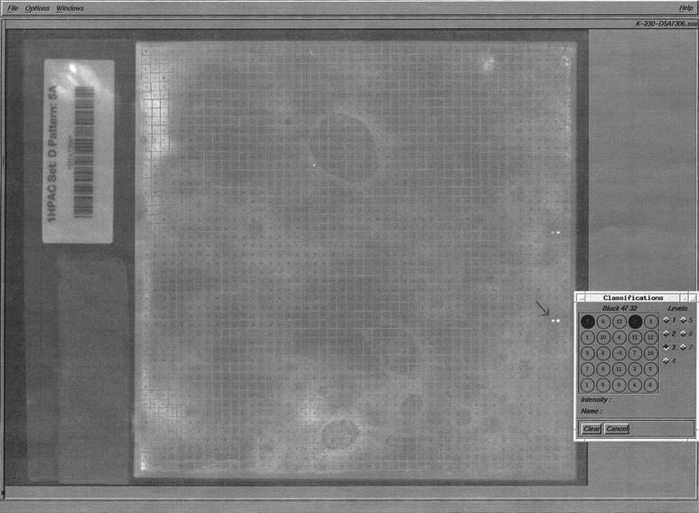
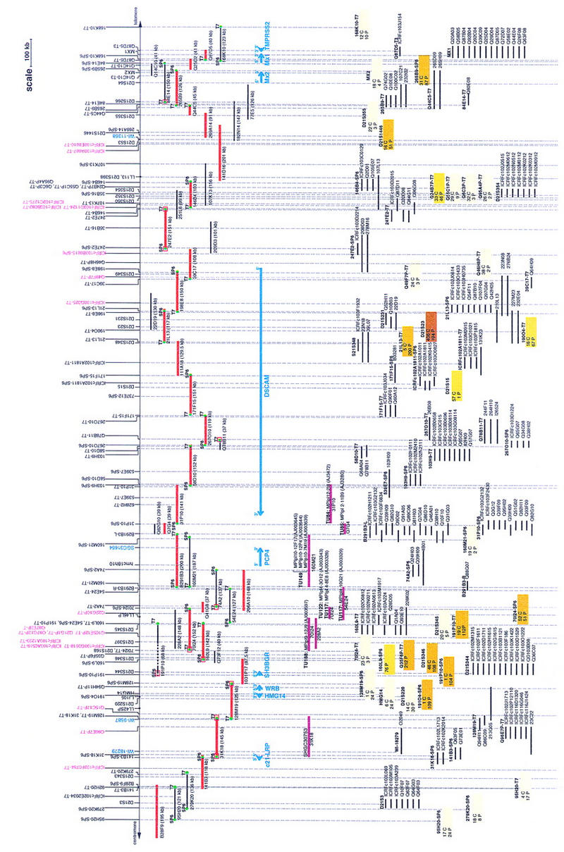
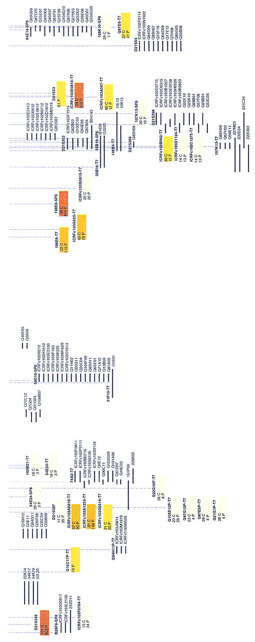
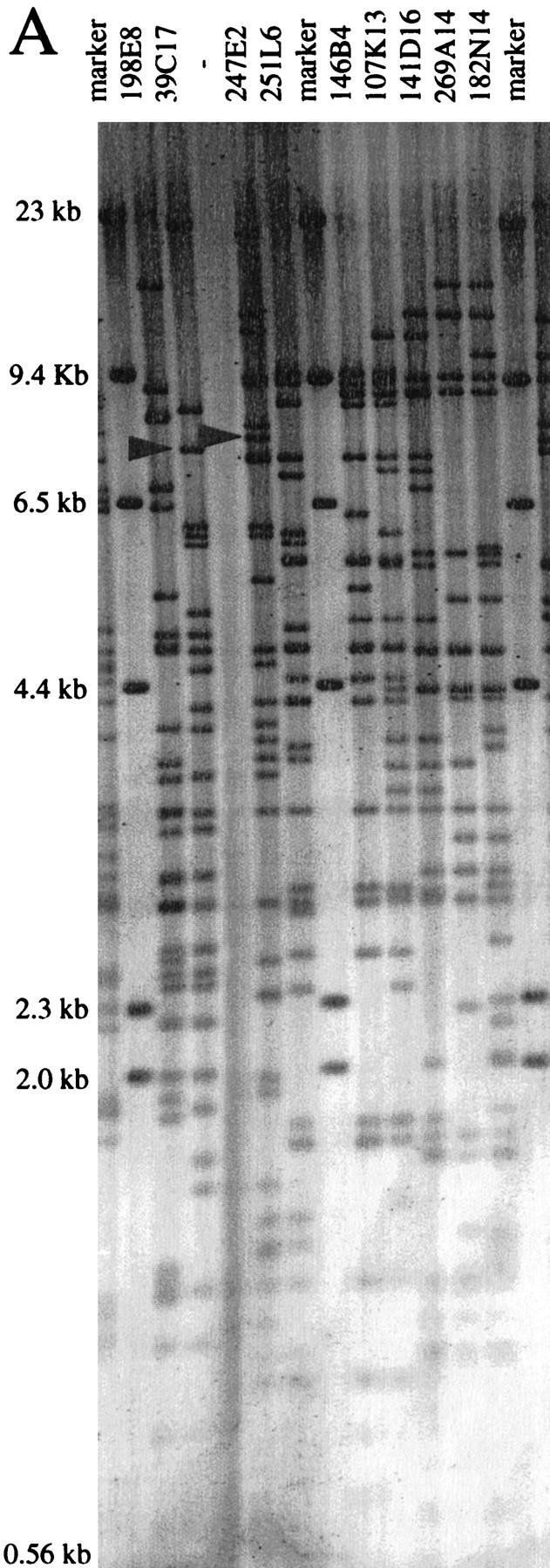
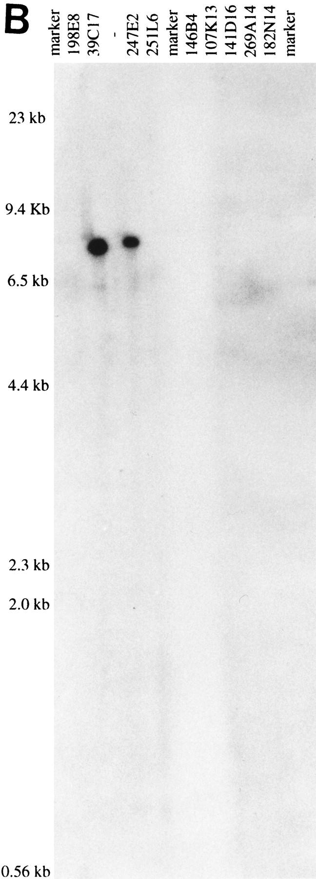
Progress in complete genomic sequencing of human chromosome 21 relies on the construction of high-quality bacterial clone maps spanning large chromosomal regions. To achieve this goal, we have applied a strategy based on nonradioactive hybridizations to contig building. A contiguous sequence-ready map was constructed in the Down syndrome congenital heart disease (DS-CHD) region in 21q22.2, as a framework for large-scale genomic sequencing and positional candidate gene approach. Contig assembly was performed essentially by high throughput nonisotopic screenings of genomic libraries, prior to clone validation by (1) restriction digest fingerprinting, (2) STS analysis, (3) Southern hybridizations, and (4) FISH analysis. The contig contains a total of 50 STSs, of which 13 were newly isolated. A minimum tiling path (MTP) was subsequently defined that consists of 20 PACs, 2 BACs, and 5 cosmids covering 3 Mb between D21S3 and MX1. Gene distribution in the region includes 9 known genes (c21-LRP, WRB, SH3BGR, HMG14, PCP4, DSCAM, MX2, MX1, and TMPRSS2) and 14 new additional gene signatures consisting of cDNA selection products and ESTs. Forthcoming genomic sequence information will unravel the structural organization of potential candidate genes involved in specific features of Down syndrome pathogenesis.
Figures
Similar articles
-
Sequence-ready BAC contig, physical, and transcriptional map of a 2-Mb region overlapping the mouse chromosome 6 host-resistance locus Cmv1.Genomics. 2000 Jun 1;66(2):161-74. doi: 10.1006/geno.2000.6186. Genomics. 2000. PMID: 10860661
-
BAC and PAC contigs covering 3.5 Mb of the Down syndrome congenital heart disease region between D21S55 and MX1 on chromosome 21.Genomics. 1997 Apr 15;41(2):218-26. doi: 10.1006/geno.1997.4657. Genomics. 1997. PMID: 9143497
-
A sequence-ready physical map of a region of 12q24.1.Genomics. 1997 Oct 15;45(2):271-8. doi: 10.1006/geno.1997.4888. Genomics. 1997. PMID: 9344649
-
Sequence-ready contig for the 1.4-cM ductal carcinoma in situ loss of heterozygosity region on chromosome 8p22-p23.Genomics. 1999 Aug 15;60(1):1-11. doi: 10.1006/geno.1999.5905. Genomics. 1999. PMID: 10458905
-
High-resolution physical map and identification of potentially regulatory sequences of the human SH3BGR located in the Down syndrome chromosomal region.Biochem Biophys Res Commun. 1997 Dec 18;241(2):321-6. doi: 10.1006/bbrc.1997.7816. Biochem Biophys Res Commun. 1997. PMID: 9425270
Cited by
-
Sequence-tagged connectors: a sequence approach to mapping and scanning the human genome.Proc Natl Acad Sci U S A. 1999 Aug 17;96(17):9739-44. doi: 10.1073/pnas.96.17.9739. Proc Natl Acad Sci U S A. 1999. PMID: 10449764 Free PMC article.
-
Structural and functional analysis of single neurons to correlate synaptic connectivity with grooming behavior.Nat Protoc. 2014 Jan;9(1):1-10. doi: 10.1038/nprot.2013.157. Epub 2013 Dec 5. Nat Protoc. 2014. PMID: 24309972
-
Overexpression of Down syndrome cell adhesion molecule impairs precise synaptic targeting.Nat Neurosci. 2013 Jun;16(6):677-82. doi: 10.1038/nn.3396. Epub 2013 May 12. Nat Neurosci. 2013. PMID: 23666178 Free PMC article.
References
-
- Chen HM, Bouras C, Antonarakis SE. Cloning of the cDNA for a human homolog of the rat pep-19 gene and mapping to chromosome 21q22.2-q22.3. Hum Genet. 1996;98:672–677. - PubMed
-
- Chumakov I, Rigault P, Guillou S, Ougen P, Billaut A, Guasconi G, Gervy P, LeGall I, Soularue P, Grinas L, et al. Continuum of overlapping clones spanning the entire human chromosome 21q. Nature. 1992;359:380–387. - PubMed
-
- Church DM, Stotler CJ, Rutter JL, Murrell JR, Trofatter JA, Buckler AJ. Isolation of genes from complex sources of mammalian genomic DNA using exon amplification. Nature Genet. 1994;6:98–105. - PubMed
Publication types
MeSH terms
LinkOut - more resources
Full Text Sources
Other Literature Sources
Miscellaneous