. 1999 Apr;103(8):1159-68.
doi: 10.1172/JCI5392.

Molecular and functional analysis of SDCT2, a novel rat sodium-dependent dicarboxylate transporter

Affiliations

Molecular and functional analysis of SDCT2, a novel rat sodium-dependent dicarboxylate transporter

X Chen et al. J Clin Invest. 1999 Apr.

Abstract

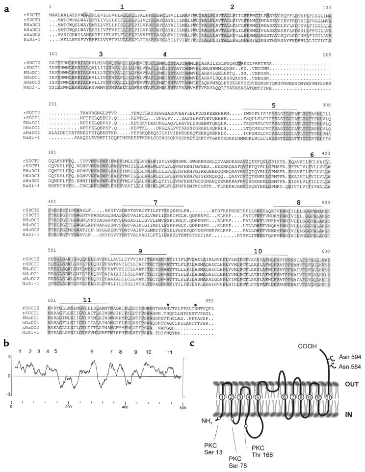
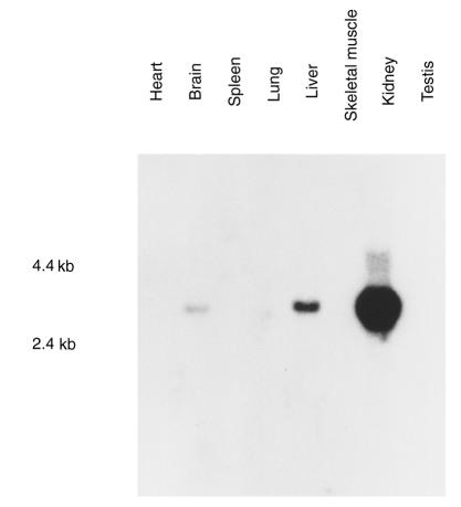
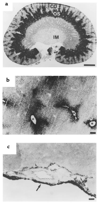
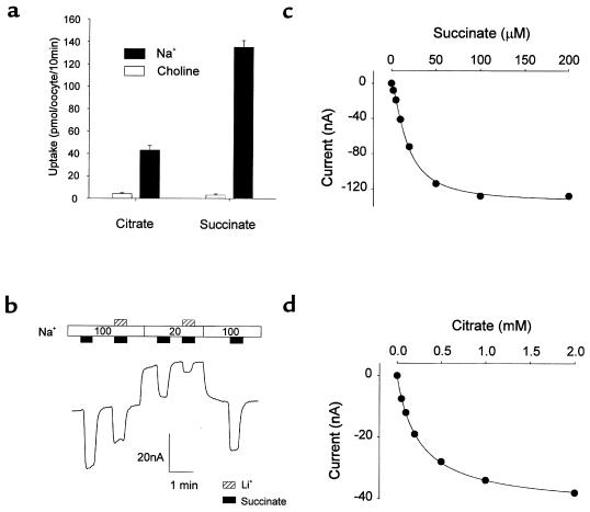
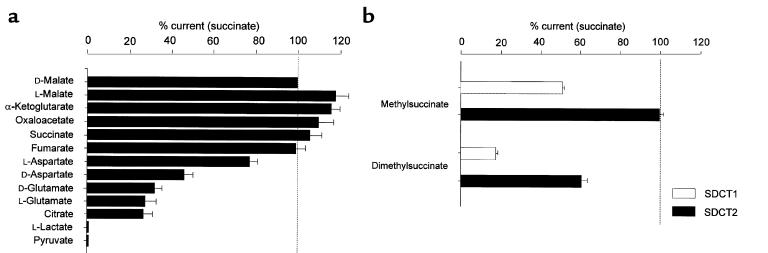
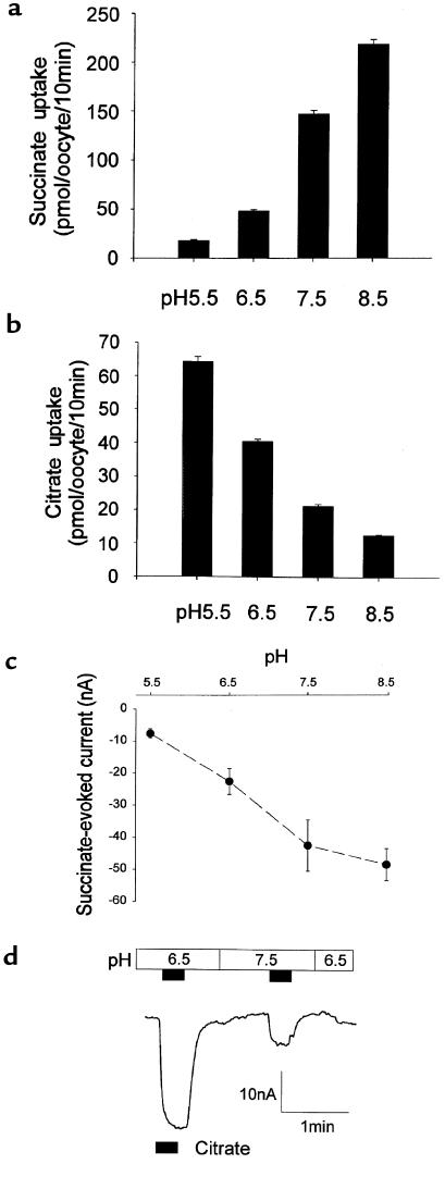
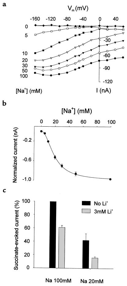
Kidney proximal tubule cells take up Krebs cycle intermediates for metabolic purposes and for secretion of organic anions through dicarboxylate/organic anion exchange. Alteration in reabsorption of citrate is closely related to renal stone formation. The presence of distinct types of sodium-coupled dicarboxylate transporters has been postulated on either side of the polarized epithelial membrane in the kidney proximal tubule. Using a PCR-based approach, we isolated a novel member of the sodium-dependent dicarboxylate/sulfate transporter called SDCT2. SDCT2 is a 600-amino acid residue protein that has 47-48% amino acid identity to SDCT1 and NaDC-1, previously identified in kidney and intestine. Northern analysis gave a single band of 3.3 kb for SDCT2 in kidney, liver, and brain. In situ hybridization revealed that SDCT2 is prominently expressed in kidney proximal tubule S3 segments and in perivenous hepatocytes, consistent with the sites of high-affinity dicarboxylate transport identified based on vesicle studies. A signal was also detected in the meningeal layers of the brain. SDCT2 expressed in Xenopus oocytes mediated sodium-dependent transport of di- and tricarboxylates with substrate preference for succinate rather than citrate, but excluding monocarboxylates. SDCT2, unlike SDCT1, displayed a unique pH dependence for succinate transport (optimal pH 7.5-8.5) and showed a high affinity for dimethylsuccinate, two features characteristic of basolateral transport. These data help to interpret the mechanisms of renal citrate transport, their alteration in pathophysiological conditions, and their role in the elimination of organic anions and therapeutic drugs.


Figures

Figure 1
Figure 1
Predicted amino acid sequence of the rat SDCT2. (a) Amino acid alignment of members of the SDCT family. Sequences of rat SDCT2 (rSDCT2; GenBank AF080451), rat SDCT1 (rSDCT1; GenBank AF058714), rabbit NaDC-1 (RNaDC-1; GenBank Q28615), human NaDC-1 (hNaDC-1; GenBank Q13183), Xenopus laevis NaDC-2 (xNaDC-2; GenBank U87318), and rat sodium-dependent sulfate transporter NaSi-1 (GenBank Q07782) are aligned by PILEUP (GCG analysis program). Identical residues are indicated by shading. The putative transmembrane domains are underlined and indicated by numbers. The consensus sequences for N-linked glycosylation (Asn 584, Asn 594) (asterisks) and potential protein kinase C phosphorylation sites (Ser 13, Ser 78, Thr 168) (boxes) are shown. (b) Hydropathy analysis of SDCT2. The hydropathy profile was analyzed by the Kyte-Doolittle algorithm with a window of 21 residues. Putative membrane-spanning domains are shown by numbers. (c) Hypothetical membrane topology model of SDCT2. Membrane-spanning domains are predicted based on the hydropathy profiles. The numbering of the putative 11 transmembrane domains corresponds to b.
Figure 2
Figure 2
Northern blot analysis of SDCT2 mRNA in rat tissues. Northern blot with poly(A)+ RNA (2 μg) from the rat tissues was hybridized with a SDCT2 cDNA probe under high-stringency conditions. The positions of size standards (measured in kb) are shown at left.
Figure 3
Figure 3
In situ hybridization of SDCT2 mRNA. Freshly frozen tissue sections (12 μm) from rats were hybridized with a digoxigenin-labeled 1.3-kb SDCT2 cRNA probe. Bright-field micrographs of sections hybridized to antisense cRNA probe are shown (ac). Hybridization of sense cRNA probe did not reveal any signals (data not shown). (a) Cross-section of kidney. A gradient of mRNA levels in S3 segment is apparent; i.e., the signal is more prominent in proximal part of the straight tubules compared with distal parts. CO, cortex; IM, inner medulla; OS, outer stripe of outer medulla. Scale bar: 2 mm. (b) Liver. SDCT2 mRNA is predominantly expressed in hepatocytes surrounding the central vein (asterisks). Scale bar: 200 μm. (c) Brain. SDCT2 mRNA is expressed by cells of meningeal layers, including dura mater (large arrow), arachnoid, and pia mater (small arrows). Scale bar: 50 μm.
Figure 4
Figure 4
Dicarboxylate transport of SDCT2 expressed in Xenopus oocytes. (a) Radiotracer uptake of succinate and citrate in SDCT2-expressing oocytes. Uptake of 100 μM [14C]succinate and 1 mM [14C]citrate was measured during 10 min in solution containing sodium or choline. (b) Time course of succinate-evoked currents. Currents were recorded at pH 7.5 and at holding membrane potential Vh = –50 mV. At 100 or 20 mM Na+, application of 50 μM succinate (filled blocks) and 3 mM Li+ (hatched blocks) is indicated. Effects of lithium were analyzed further in Figure 7b. (c) Concentration dependence of SDCT2-mediated currents. Currents were measured at different concentrations of succinate (0–200 μM, pH 7.5) and at Vh = –50 mV. (d) Concentration dependence of citrate-evoked currents. Currents were measured at different concentrations of citrate (0–2.0 mM, pH 7.5) and at Vh = –50 mV.
Figure 5
Figure 5
Substrate specificity of SDCT2. (a) Currents induced by 1 mM substrate are shown as a percentage of those evoked by succinate (n = 3–6). Currents generated by different dicarboxylates were in the following order: D-malate, L-malate, α-ketoglutarate, oxaloacetate, succinate, fumarate. No measurable currents were detected for monocarboxylates. (b) Comparison of transport selectivity of SDCT1 and SDCT2. Currents evoked by DL-methylsuccinate and 2,3,-dimethylsuccinate (50 μM) were measured in the oocytes expressing either SDCT1 (hatched bars) or SDCT2 (filled bars). The currents are indicated as a percentage of the succinate-evoked current (50 μM) observed in the same oocyte (n = 3–4).
Figure 6
Figure 6
pH effects on succinate and citrate transport. Proton dependency of uptake for succinate (a) and citrate (b). Uptake of 100 μM [14C]succinate or 1 mM [14C]citrate was measured during 10 min under indicated pH. (c) pH dependency of succinate-evoked current. Average currents evoked by succinate (20 μM) at four different pH values from 5.5 to 8.5 were measured (Vh = –50 mV, n = 3). (d) pH dependency of citrate-evoked current. Inward currents generated by citrate (200 μM) are demonstrated by superfusing oocytes with solutions at pH 6.5 and pH 7.5, respectively (Vh = –50 mV). Citrate addition (filled blocks) is indicated.
Figure 7
Figure 7
Cationic effects on succinate-evoked currents. (a) Current-voltage relationship under various sodium concentrations. Under different sodium concentrations (0–100 mM), steady-state current and voltage relation between –160 to + 60 mV were obtained by subtracting currents in the presence and absence of 100 μM succinate. (b) Sigmoidal relationship of currents versus sodium concentration. At Vh = –50 mV, currents evoked by 100 μM succinate at different sodium concentrations were normalized by those at 100 mM NaCl medium. The curve was fitted by the Hill equation. (c) Effects of lithium on SDCT2 currents. Succinate-evoked currents (at 50 μM) in the presence or absence of lithium are shown as a percentage of those at 100 mM NaCl medium without Li+. In absence of Li+, average currents at 20 mM Na+ are 42 ± 10% of those at 100 mM Na+.

Similar articles

Cited by

References

    1. Murer, H., Manganel, M., and Roch-Ramel, F. 1992. Tubular transport of monocarboxylates, Krebs cycle intermediates, and inorganic sulfate. In Handbook of physiology: renal physiology. E.E. Windhager, editor. American Physiological Society. Bethesda, MD. 2165–2188.
    1. Hamm LL. Renal handling of citrate. Kidney Int. 1990;38:728–735. - PubMed
    1. Brennan S, Hering-Smith K, Hamm LL. Effect of pH on citrate reabsorption in the proximal convoluted tubule. Am J Physiol. 1988;255:F301–F306. - PubMed
    1. Wright EM. Transport of carboxylic acids by renal membrane vesicles. Annu Rev Physiol. 1985;47:127–141. - PubMed
    1. Wright SH, Kippen I, Wright EM. Effect of pH on the transport of Krebs cycle intermediates in renal brush border membranes. Biochim Biophys Acta. 1982;684:287–290. - PubMed

Publication types

Associated data