

Perforin-independent beta-cell destruction by diabetogenic CD8(+) T lymphocytes in transgenic nonobese diabetic mice
- PMID: 10207172
- PMCID: PMC408282
- DOI: 10.1172/JCI6266
Perforin-independent beta-cell destruction by diabetogenic CD8(+) T lymphocytes in transgenic nonobese diabetic mice
Abstract
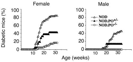
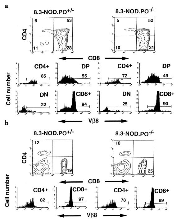
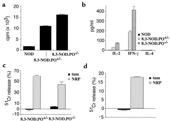
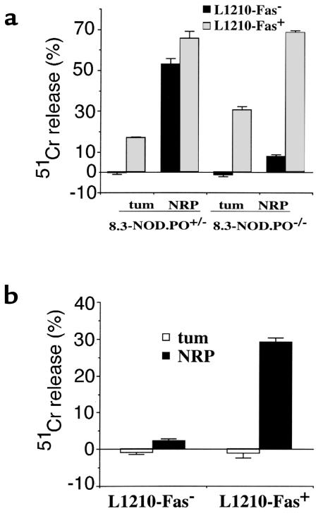
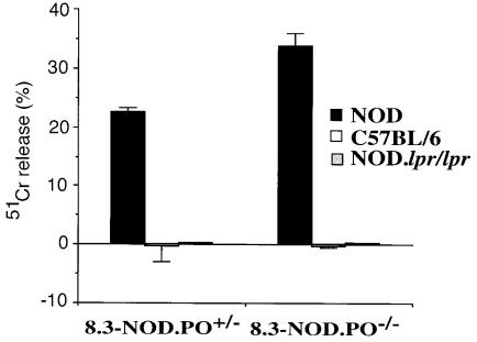
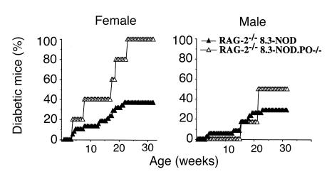
Autoimmune diabetes in nonobese diabetic (NOD) mice results from destruction of pancreatic beta cells by T lymphocytes. It is believed that CD8(+) cytotoxic T lymphocytes (CTLs) effect the initial beta-cell insult in diabetes, but the mechanisms remain unclear. Studies of NOD.lpr mice have suggested that disease initiation is a Fas-dependent process, yet perforin-deficient NOD mice rarely develop diabetes despite expressing Fas. Here, we have investigated the role of perforin and Fas in the ability of beta cell-reactive CD8(+) T cells bearing a T-cell receptor (8.3-TCR) that is representative of TCRs used by CD8(+) CTLs propagated from the earliest insulitic lesions of NOD mice, and that targets an immunodominant peptide/H-2Kd complex on beta cells, to effect beta-cell damage in vitro and in vivo. In vitro, 8.3-CTLs killed antigenic peptide-pulsed non-beta-cell targets via both perforin and Fas, but they killed NOD beta cells via Fas exclusively. Perforin-deficient 8.3-TCR-transgenic NOD mice expressing an oligoclonal or monoclonal T-cell repertoire developed diabetes even more frequently than their perforin-competent littermates. These results demonstrate that diabetogenic CD8(+) CTLs representative of CTLs putatively involved in the initiation of autoimmune diabetes kill beta cells in a Fas-dependent and perforin-independent manner.
Figures

Similar articles
-
IL-1alpha, IL-1beta, and IFN-gamma mark beta cells for Fas-dependent destruction by diabetogenic CD4(+) T lymphocytes.J Clin Invest. 2000 Feb;105(4):459-68. doi: 10.1172/JCI8185. J Clin Invest. 2000. PMID: 10683375 Free PMC article.
-
Comparing the relative role of perforin/granzyme versus Fas/Fas ligand cytotoxic pathways in CD8+ T cell-mediated insulin-dependent diabetes mellitus.J Immunol. 1999 Oct 15;163(8):4335-41. J Immunol. 1999. PMID: 10510373
-
TNF receptor 1-dependent beta cell toxicity as an effector pathway in autoimmune diabetes.J Immunol. 1999 Apr 15;162(8):4598-605. J Immunol. 1999. PMID: 10201999
-
Beta cell destruction in the development of autoimmune diabetes in the non-obese diabetic (NOD) mouse.Diabetes Metab Res Rev. 2000 Jul-Aug;16(4):251-61. doi: 10.1002/1520-7560(200007/08)16:4<251::aid-dmrr126>3.0.co;2-c. Diabetes Metab Res Rev. 2000. PMID: 10934453 Review.
-
Cellular and molecular pathogenic mechanisms of insulin-dependent diabetes mellitus.Ann N Y Acad Sci. 2001 Apr;928:200-11. doi: 10.1111/j.1749-6632.2001.tb05650.x. Ann N Y Acad Sci. 2001. PMID: 11795511 Review.
Cited by
-
B-cell cross-presentation of autologous antigen precipitates diabetes.Diabetes. 2012 Nov;61(11):2893-905. doi: 10.2337/db12-0006. Epub 2012 Jul 24. Diabetes. 2012. PMID: 22829452 Free PMC article.
-
Fas/Fas-Ligand Interaction As a Mechanism of Immune Homeostasis and β-Cell Cytotoxicity: Enforcement Rather Than Neutralization for Treatment of Type 1 Diabetes.Front Immunol. 2017 Mar 27;8:342. doi: 10.3389/fimmu.2017.00342. eCollection 2017. Front Immunol. 2017. PMID: 28396667 Free PMC article. No abstract available.
-
Effector lymphocytes in islet cell autoimmunity.Rev Endocr Metab Disord. 2003 Sep;4(3):271-80. doi: 10.1023/a:1025156413404. Rev Endocr Metab Disord. 2003. PMID: 14501178 Review. No abstract available.
-
Prospects for the prevention and reversal of type 1 diabetes mellitus.Drugs. 2002;62(18):2617-35. doi: 10.2165/00003495-200262180-00005. Drugs. 2002. PMID: 12466001 Review.
-
IL-1alpha, IL-1beta, and IFN-gamma mark beta cells for Fas-dependent destruction by diabetogenic CD4(+) T lymphocytes.J Clin Invest. 2000 Feb;105(4):459-68. doi: 10.1172/JCI8185. J Clin Invest. 2000. PMID: 10683375 Free PMC article.
References
-
- Delovitch T, Singh B. The nonobese diabetic mouse as a model of autoimmune diabetes: immune disregulation gets the NOD. Immunity. 1997;7:727–738. - PubMed
-
- Tisch R, McDevitt H. Insulin-dependent diabetes mellitus. Cell. 1996;85:291–297. - PubMed
-
- Miller BJ, Appel MC, O’Neil JJ, Wicker LS. Both the Lyt-2+ and L3T4+ T cell subsets are required for the transfer of diabetes in non-obese diabetic mice. J Immunol. 1988;140:52–58. - PubMed
-
- Yagi H, et al. Analysis of the roles of CD4+ and CD8+ T cells in autoimmune diabetes of NOD mice using transfer to NOD athymic nude mice. Eur J Immunol. 1992;22:2387–2393. - PubMed
Publication types
MeSH terms
Substances
LinkOut - more resources
Full Text Sources
Other Literature Sources
Medical
Molecular Biology Databases
Research Materials
Miscellaneous
