Homozygosity mapping and linkage analysis demonstrate that autosomal recessive congenital hereditary endothelial dystrophy (CHED) and autosomal dominant CHED are genetically distinct
- PMID: 10209448
- PMCID: PMC1722772
- DOI: 10.1136/bjo.83.1.115
Homozygosity mapping and linkage analysis demonstrate that autosomal recessive congenital hereditary endothelial dystrophy (CHED) and autosomal dominant CHED are genetically distinct
Abstract
Background: Congenital hereditary endothelial dystrophy (CHED) is a corneal dystrophy characterised by diffuse bilateral corneal clouding resulting in impaired vision. It is inherited in either an autosomal dominant (AD) or autosomal recessive (AR) manner. The AD form of CHED has been mapped to the pericentromeric region of chromosome 20. Another endothelial dystrophy, posterior polymorphous dystrophy (PPM), has been linked to a larger but overlapping region on chromosome 20. A large, Irish, consanguineous family with AR CHED was investigated to determine if there was linkage to this region.
Methods: The technique of linkage analysis with polymorphic microsatellite markers amplified by polymerase chain reaction (PCR) was used. In addition, a DNA pooling approach to homozygosity mapping was employed to demonstrate the efficiency of this method.
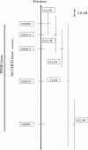
Results: Conventional genetic analysis in addition to a pooled DNA strategy excludes linkage of AR CHED to the AD CHED and larger PPMD loci.
Conclusion: This demonstrates that AR CHED is genetically distinct from AD CHED and PPMD.
Figures


Similar articles
-
Genetics of the corneal endothelial dystrophies: an evidence-based review.Clin Genet. 2013 Aug;84(2):109-19. doi: 10.1111/cge.12191. Epub 2013 Jun 10. Clin Genet. 2013. PMID: 23662738 Free PMC article. Review.
-
Localization of the gene for autosomal recessive congenital hereditary endothelial dystrophy (CHED2) to chromosome 20 by homozygosity mapping.Genomics. 1999 Oct 1;61(1):1-4. doi: 10.1006/geno.1999.5920. Genomics. 1999. PMID: 10512674
-
Exclusion of AR-CHED from the chromosome 20 region containing the PPMD and AD-CHED loci.Ophthalmic Genet. 1999 Dec;20(4):243-9. doi: 10.1076/opge.20.4.243.2273. Ophthalmic Genet. 1999. PMID: 10617922
-
Linkage of congenital hereditary endothelial dystrophy to chromosome 20.Hum Mol Genet. 1995 Dec;4(12):2395-8. doi: 10.1093/hmg/4.12.2395. Hum Mol Genet. 1995. PMID: 8634716
-
Congenital hereditary endothelial dystrophy with progressive sensorineural deafness (Harboyan syndrome).Orphanet J Rare Dis. 2008 Oct 15;3:28. doi: 10.1186/1750-1172-3-28. Orphanet J Rare Dis. 2008. PMID: 18922146 Free PMC article. Review.
Cited by
-
Identification and in silico analysis of a spectrum of SLC4A11 variations in Indian familial and sporadic cases of congenital hereditary endothelial dystrophy.Orphanet J Rare Dis. 2022 Sep 17;17(1):361. doi: 10.1186/s13023-022-02521-4. Orphanet J Rare Dis. 2022. PMID: 36115991 Free PMC article.
-
Updates on congenital hereditary endothelial dystrophy.Taiwan J Ophthalmol. 2023 Nov 28;13(4):405-416. doi: 10.4103/tjo.TJO-D-23-00135. eCollection 2023 Oct-Dec. Taiwan J Ophthalmol. 2023. PMID: 38249503 Free PMC article. Review.
-
Genetics of corneal endothelial dystrophies.J Genet. 2009 Dec;88(4):487-94. doi: 10.1007/s12041-009-0067-1. J Genet. 2009. PMID: 20090209 Review.
-
The IC3D classification of the corneal dystrophies.Cornea. 2008 Dec;27 Suppl 2(Suppl 2):S1-83. doi: 10.1097/ICO.0b013e31817780fb. Cornea. 2008. PMID: 19337156 Free PMC article. Review.
-
Genetics of the corneal endothelial dystrophies: an evidence-based review.Clin Genet. 2013 Aug;84(2):109-19. doi: 10.1111/cge.12191. Epub 2013 Jun 10. Clin Genet. 2013. PMID: 23662738 Free PMC article. Review.
References
MeSH terms
LinkOut - more resources
Full Text Sources
Research Materials
