. 1999 Apr;116(1):48-56.
doi: 10.1046/j.1365-2249.1999.00857.x.

Bladder cancer immunogenicity: expression of CD80 and CD86 is insufficient to allow primary CD4+ T cell activation in vitro

Affiliations

Bladder cancer immunogenicity: expression of CD80 and CD86 is insufficient to allow primary CD4+ T cell activation in vitro

S J Pettit et al. Clin Exp Immunol. 1999 Apr.

Abstract

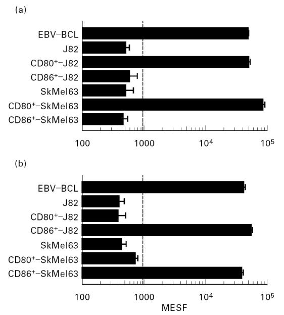
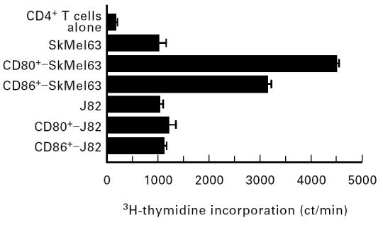
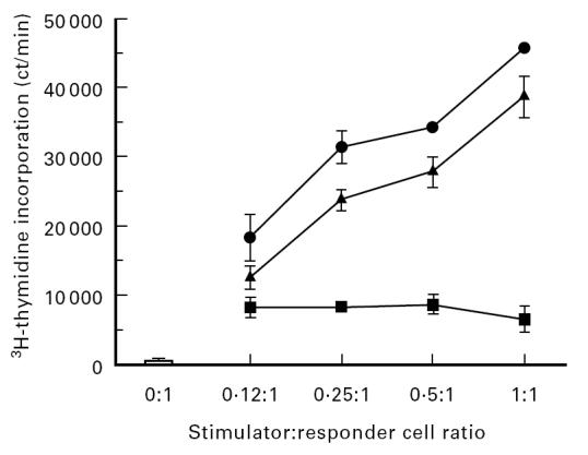
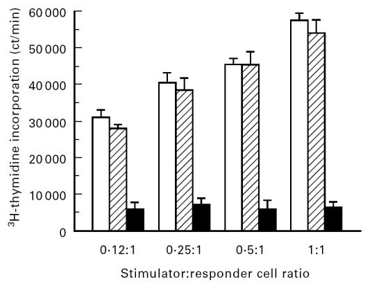
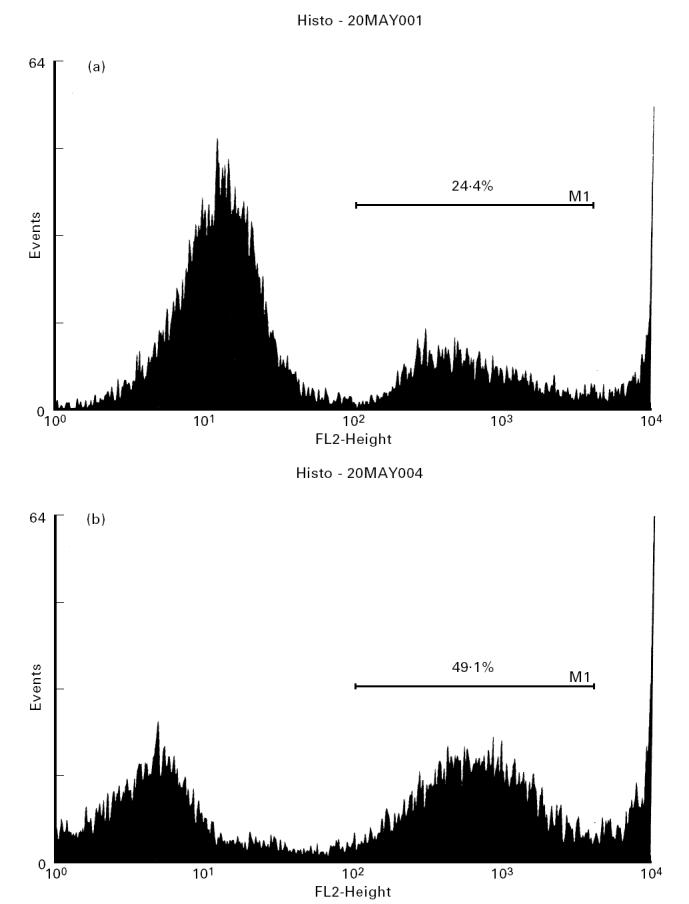
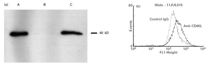
Transitional cell carcinomas (TCC) of the urinary bladder are known to express proteins which can yield potentially immunogenic peptide epitopes for expression in the context of cell surface class I or class II MHC antigens. However, additional costimulatory ligands must also be expressed before such a cell might directly induce full activation and proliferation of resting, antigen-specific T lymphocytes. Intravesical therapy might be used to manipulate T cell costimulation in order to promote specific rejection of TCC cells. This in vitro study examined the potential of such a strategy by transfection of the prototypical TCC line J82 with the important costimulatory molecules CD80 (B7-1) and CD86 (B7-2). Untransfected J82 cells expressed class I and II MHC antigens, a range of cell adhesion molecules, though did not induce T cell proliferation in a robust, allogeneic co-culture system. Transfected J82 cells expressed CD80 or CD86 at levels comparable to an antigen-presenting B cell line. Furthermore, functional surface expression of CD80 and CD86 was demonstrated in a mitogen-dependent assay of costimulation. However, neither CD80+ nor CD86+ transfectant J82 cells could induce significant proliferation of antigen-specific CD4+ T cells. Further analysis showed that bystander J82 cells could inhibit independent T cell activation in an effect dependent on direct cell contact. This inhibitory effect was associated with increased cell death in the responding lymphocyte population and is concordant with surface expression of CD95L by the J82 cell line.


Figures

Fig 1
Fig 1
Representative results showing expression of (a) CD80 and (b) CD86 on the surface of the Epstein–Barr virus-transformed B cell lines (EBV-BCL), untransfected and transfected SkMel63 lines, and untransfected and transfected J82 lines. Each point represents the mean of duplicate determinations; error bars show the s.e.m.; the dotted line represents the upper 95% confidence interval (CI) of negative control staining. MESF, Median equivalent molecules of soluble fluorescein.
Fig 2
Fig 2
Representative results showing proliferation of CD4+ T cells in co-culture with untransfected, CD80+ and CD86+ IFN-γ-pretreated J82 lines and SkMel63 lines at an optimal stimulator:responder cell ratio of 0.25:1. Proliferation was measured on day 5 by 3H-thymidine incorporation, and expressed as ct/min. Each point represents the mean of triplicate determinations, error bars show the s.e.m.
Fig 3
Fig 3
Representative results showing proliferation of phytohaemagglutinin (PHA)-stimulated CD4+ T cells alone (□) and in co-culture with J82 cells (▪), CD80+-J82 cells (▴) and CD86+-J82 cells (•). Proliferation was measured on day 3 by 3H-thymidine incorporation, and expressed as ct/min. Each point represents the mean of triplicate determinations, error bars show the s.e.m.
Fig 4
Fig 4
Representative results showing proliferation of an Epstein–Barr virus-transformed B cell line (EBV-BCL)-stimulated mixed lymphocyte reaction (MLR) within inserts above culture medium (□), within inserts above a J82 monolayer (hatched bars), and placed directly upon a J82 monolayer (▪). Proliferation was measured on day 5 by 3H-thymidine incorporation, and expressed as ct/min. Each point represents the mean of duplicate determinations, error bars show the s.e.m.
Fig 5
Fig 5
DNA fluorescence flow cytometric profiles of propidium iodide (PI)-stained cell populations harvested from a positive control mixed lymphocyte reaction (MLR) (a) and an MLR generated in direct contact with a J82 monolayer (b). The gate M1 encloses the PI-stained DNA peak for analysis. Representative results from a single experiment are given.
Fig 6
Fig 6
Representative results showing expression of CD95L. (a) Western blot demonstrating expression of the 40-kD CD95L protein by SW620 (lane A) and J82 (lane C) lines, but not by the SkMel63 line (lane B). (b) Flow cytometric histograms demonstrating surface expression of CD95L on J82 cells stained with an anti-CD95L or an isotype-matched control antibody.

Similar articles

Cited by

References

    1. Knight SC, Stagg AJ. Antigen presenting cell types. Curr Opin Immunol. 1993;5:374–82. - PubMed
    1. Lenschow DJ, Walunas TL, Bluestone JA. CD28/B7 system of T-cell costimulation. Ann Rev Immunol. 1996;14:233–58. - PubMed
    1. Hart I, Colaco C. Fusion induces tumour rejection. Nature. 1997;388:626–7. - PubMed
    1. Boon T, van der Bruggen P. Human tumor antigens recognized by T lymphocytes. J Exp Med. 1996;183:725–9. - PMC - PubMed
    1. Ostrand Rosenberg S. Tumor immunotherapy—the tumor cell as an antigen presenting cell. Curr Opin Immunol. 1994;6:722–7. - PubMed

Publication types

MeSH terms