

T cell responses to the putative dominant autoepitope in primary biliary cirrhosis (PBC)
- PMID: 10209517
- PMCID: PMC1905223
- DOI: 10.1046/j.1365-2249.1999.00803.x
T cell responses to the putative dominant autoepitope in primary biliary cirrhosis (PBC)
Abstract
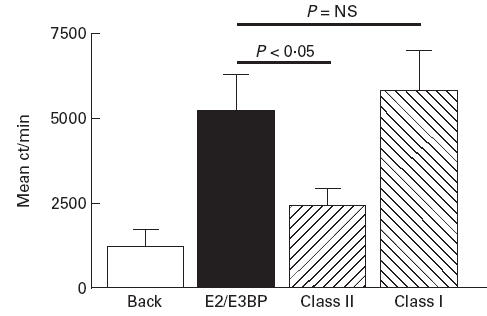
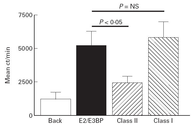
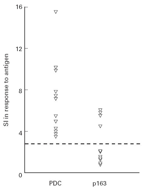
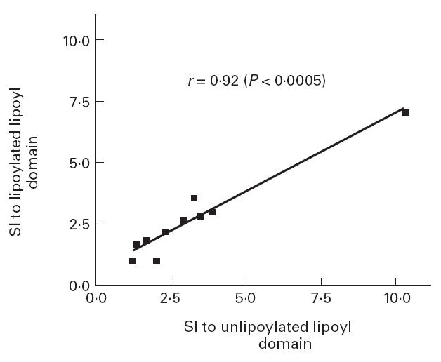
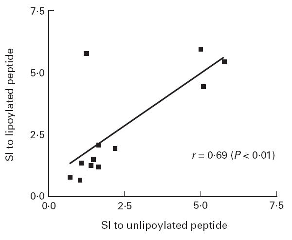
PBC is characterized by T cell-mediated destruction of the biliary epithelial cells lining the small intrahepatic bile ducts. The E2 and E3 binding protein (E3BP (protein X)) components of pyruvate dehydrogenase complex (PDC) are disease-specific autoantigens in PBC. Attempts to localize the T cell autoepitopes within PDC-E2 have, however, generated contradictory results. One study has suggested the presence of T cell epitopes throughout PDC-E2, whilst another has identified a single dominant 14 amino acid T cell epitope (p163) spanning the lipoic acid binding lysine residue in the inner lipoyl domain (ILD) of PDC-E2. The aim of the current study was to determine the prevalence of T cell responses to p163 and PDC-E2 ILD, and the role played by lipoylation of these antigens in their immunogenicity, in a UK PBC population. We found that the majority of the PBC patients showing a 6-day peripheral blood T cell proliferative response to native human PDC also responded, in a MHC class II-restricted fashion, to biochemically purified PDC-E2 and E3BP (which co-purify) (9/10 positive (SI > 2.76), mean SI 5.74 +/- 5.04 (PDC-E2/E3BP) versus 6.67 +/- 3.84 (PDC), P = NS), implying that the important PBC-specific T cell epitopes are contained within the PDC-E2 or E3BP components of PDC. Only a minority of patients responsive to PDC, however, responded to either lipoylated recombinant PDC-E2 ILD (4/10 positive, mean SI 1.98 +/- 1.24, P < 0.005 versus PDC response) or lipoylated p163 (4/12 positive, mean SI 1.90 +/- 1.58, P < 0.001). The lipoylation state did not affect the T cell response to either ILD or p163. Our findings suggest that in some UK patients with PBC there are immunodominant T cell autoepitopes within PDC-E2/E3BP which are outside the ILD of PDC-E2.
Figures

References
-
- Kaplan M. Primary biliary cirrhosis. N Engl J Med. 1996;335:1570–9. - PubMed
-
- Jones DEJ, Bassendine MF. Primary biliary cirrhosis. J Int Med. 1997;241:127–30. - PubMed
-
- Bjorkland A, Festin R, Mendel-Hartvig I, et al. Blood and liver infiltrating lymphocytes in primary biliary cirrhosis: increase in activated T and natural killer cells and recruitment of primed memory T-cells. Hepatology. 1991;13:1106–11. - PubMed
Publication types
MeSH terms
Substances
Grants and funding
LinkOut - more resources
Full Text Sources
Research Materials
Miscellaneous
