

Increased bcl-2 expression in lymphocytes and its association with hepatocellular damage in patients with autoimmune hepatitis
- PMID: 10209518
- PMCID: PMC1905214
- DOI: 10.1046/j.1365-2249.1999.00861.x
Increased bcl-2 expression in lymphocytes and its association with hepatocellular damage in patients with autoimmune hepatitis
Abstract
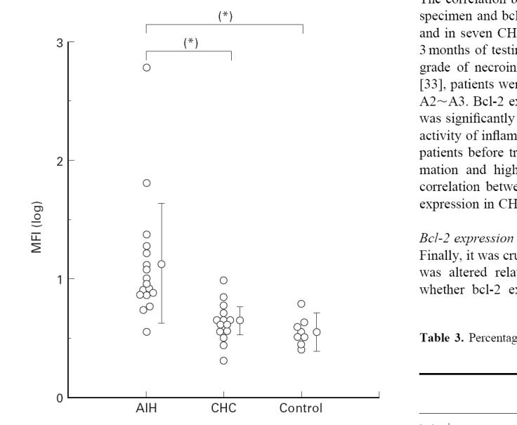
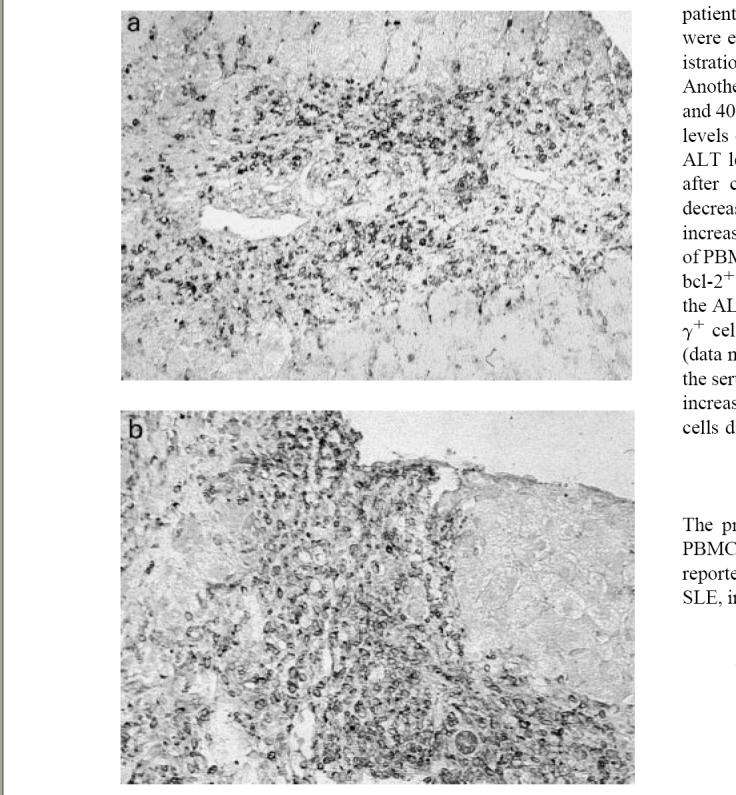
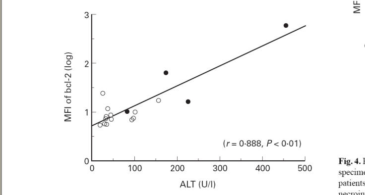
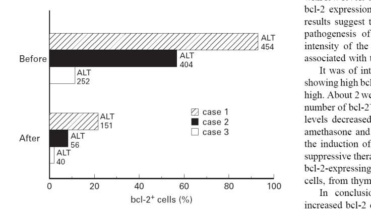
The proto-oncogene product bcl-2 is known to inhibit apoptotic cell death, and its dysregulation might play a critical role in the development of autoimmune disease. To elucidate the role of bcl-2 in autoimmune hepatitis (AIH), its expression in peripheral blood mononuclear cells (PBMC) and in liver-infiltrating lymphocytes (LIL) was investigated. Increased bcl-2 expression in PBMC was found in AIH patients compared with that in chronic hepatitis C (CHC) patients and in healthy controls. The level of bcl-2 expression significantly correlated with serum ALT level. Further analysis showed that CD4+ T cells are enriched in bcl-2-expressing PBMC. To characterize the Th1/Th2 profile of bcl-2-expressing CD4+ T cells, intracellular interferon-gamma (IFN-gamma) and IL-4 were analysed. The results revealed that most of the bcl-2-expressing cells were found to be IFN-gamma-secreting Th1 cells. In three patients for whom their clinical courses could be followed, bcl-2 expression was decreased after the initiation of immunosuppressive therapy with corticosteroids. However, the level of IFN-gamma + cells was not altered. Immunohistochemical analysis also showed that large amounts of bcl-2+ cells were observed in periportal area in the liver. In conclusion, bcl-2-expressing cells were shown to be increased in peripheral blood and liver in AIH and the bcl-2 product was expressed mainly in CD4+ Th1-type cells, suggesting that these cells might promote the cellular immune response and contribute to the development of hepatitis and hepatocellular damage in AIH.
Figures

Similar articles
-
Analysis of CD28 and bcl-2 expression on peripheral blood and liver-infiltrating mononuclear cells in patients with autoimmune hepatitis.J Clin Immunol. 2006 Jul;26(4):323-30. doi: 10.1007/s10875-006-9030-6. Epub 2006 Jun 16. J Clin Immunol. 2006. PMID: 16779679
-
Increase in CD95 (Fas/APO-1)-positive CD4+ and CD8+ T cells in peripheral blood derived from patients with autoimmune hepatitis or chronic hepatitis C with autoimmune phenomena.J Gastroenterol Hepatol. 2000 Jan;15(1):69-75. doi: 10.1046/j.1440-1746.2000.02044.x. J Gastroenterol Hepatol. 2000. PMID: 10719750
-
Bcl-2 and Fas expression in peripheral blood leukocytes of patients with alcoholic and autoimmune liver disorders.Hum Exp Toxicol. 2016 Aug;35(8):799-807. doi: 10.1177/0960327115607078. Epub 2015 Oct 1. Hum Exp Toxicol. 2016. PMID: 26429926
-
Aetiopathogenesis of autoimmune hepatitis.World J Gastroenterol. 2008 Jun 7;14(21):3306-12. doi: 10.3748/wjg.14.3306. World J Gastroenterol. 2008. PMID: 18528928 Free PMC article. Review.
-
[Immunopathology of chronic liver diseases].Verh Dtsch Ges Pathol. 1995;79:186-97. Verh Dtsch Ges Pathol. 1995. PMID: 8600684 Review. German.
Cited by
-
Usefulness of liver infiltrating CD86-positive mononuclear cells for diagnosis of autoimmune hepatitis.World J Gastroenterol. 2006 Apr 28;12(16):2523-9. doi: 10.3748/wjg.v12.i16.2523. World J Gastroenterol. 2006. PMID: 16688797 Free PMC article.
-
Melanoma Clinical Decision Support System: An Artificial Intelligence-Based Tool to Diagnose and Predict Disease Outcome in Early-Stage Melanoma Patients.Cancers (Basel). 2023 Apr 6;15(7):2174. doi: 10.3390/cancers15072174. Cancers (Basel). 2023. PMID: 37046835 Free PMC article.
-
Analysis of CD28 and bcl-2 expression on peripheral blood and liver-infiltrating mononuclear cells in patients with autoimmune hepatitis.J Clin Immunol. 2006 Jul;26(4):323-30. doi: 10.1007/s10875-006-9030-6. Epub 2006 Jun 16. J Clin Immunol. 2006. PMID: 16779679
-
Chinese medicine bu xu hua yu recipe for the regulation of treg/th17 ratio imbalance in autoimmune hepatitis.Evid Based Complement Alternat Med. 2015;2015:461294. doi: 10.1155/2015/461294. Epub 2015 Apr 21. Evid Based Complement Alternat Med. 2015. PMID: 25977698 Free PMC article.
-
Baicalein selectively induces apoptosis in activated lymphocytes and ameliorates concanavalin a-induced hepatitis in mice.PLoS One. 2013 Jul 22;8(7):e69592. doi: 10.1371/journal.pone.0069592. Print 2013. PLoS One. 2013. PMID: 23894507 Free PMC article.
References
-
- Patel T, Gores GJ. Apoptosis and hepatobiliary disease. Hepatol. 1995;21:1725–41. - PubMed
-
- Feldmann G. Liver apoptosis. J Hepatol. 1997;26:1–11. - PubMed
-
- Krammer PK, Behrmann I, Daniel P, Dhein J, Debatin K-M. Regulation of apoptosis in the immune system. Curr Opin Immunol. 1994;6:279–89. - PubMed
-
- Schwartz LM, Osborne BA. Programmed cell death, apoptosis and killer genes. Immunol Today. 1993;14:582–90. - PubMed
-
- Jenkinson EJ, Kingston R, Smith CA, Williams GT, Owen JJT. Antigen-induced apoptosis in developing T cells: a mechanism for negative selection of the T cell receptor repertoire. Eur J Immunol. 1989;19:2175–7. - PubMed
MeSH terms
Substances
LinkOut - more resources
Full Text Sources
Other Literature Sources
Research Materials
