. 1999 Apr;116(1):181-7.
doi: 10.1046/j.1365-2249.1999.00834.x.

Active suppression in orally tolerized rats coincides with in situ transforming growth factor-beta (TGF-beta) expression in the draining lymph nodes

Affiliations

Active suppression in orally tolerized rats coincides with in situ transforming growth factor-beta (TGF-beta) expression in the draining lymph nodes

B S Lundin et al. Clin Exp Immunol. 1999 Apr.

Abstract

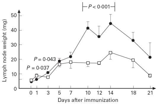
Adult rats were fed pellets containing ovalbumin (OvA) during 4 weeks, and were 2 weeks thereafter immunized subcutaneously with a mixture of OvA and human serum albumin (HSA) in Freund's complete adjuvant (day 0). As a result of the immunization, the draining lymph nodes of the nontolerized (control) rats were heavily enlarged from day 10 to day 18; however, this size increase was absent in the OvA-fed rats. This manifestation of active suppression in the tolerized rats was preceded by the appearance of scattered CD4+ TGF-beta-expressing T cells in the T cell area of their lymph nodes (days 5-8); correspondingly, the levels of TGF-beta mRNA in the nodes were elevated in the tolerant rats compared with the control rats. The anti-OvA antibody levels in sera from the rats revealed that there was an initial B cell priming in the OvA-fed group, with levels higher than in the control group during the first week. Thereafter, suppression governed the response, and from day 10 onwards the anti-OvA levels were considerably lower than in the controls. When other groups of animals were pretreated with neutralizing anti-TGF-beta antibodies 1 day before the immunization, the anti-OvA response of the OvA-fed rats was restored to the levels of the control group, demonstrating the importance of TGF-beta in the maintenance of suppression. In conclusion, we demonstrate that TGF-beta-producing cells appear in the draining lymph nodes shortly after immunization in rats made orally tolerant using a relatively high-dose feeding regime; these cells are probably responsible for the down-regulation of the immune response observed in the OvA-fed rats.


Figures

Fig 1
Fig 1
Rats made tolerant to ovalbumin (OvA) (□), and control rats (•), were immunized subcutaneously in the hind leg with a mixture of OvA and human serum albumin (HSA) in Freund's complete adjuvant. The serum antibody response to OvA (a) and HSA (b) was followed. Three animals from each group were killed at every time point, and the results are shown as mean ± s.d. The antibody levels were compared using the Mann–Whitney test.
Fig 2
Fig 2
Rats made tolerant to ovalbumin (OvA) (□), and control rats (•), were immunized subcutaneously in the hind leg with a mixture of OvA and human serum albumin (HSA) in Freund's complete adjuvant. At different intervals after challenge, the draining lymph nodes were removed and weighed. Three animals from each group were killed at every time point. The results are shown as mean ± s.e.m.; three experiments with similar results were performed. The weights were compared using the Mann–Whitney test.
Fig 3
Fig 3
Rats made tolerant to ovalbumin (OvA) (□), and control rats (▪), were immunized subcutaneously in the hind leg with a mixture of OvA and human serum albumin (HSA) in Freund's complete adjuvant. At different intervals after challenge, the draining lymph nodes were removed and TGF-β-expressing cells were enumerated, using immunohistochemistry. Six to nine animals from each group were killed during each time interval, and four different sections from each rat were evaluated. The results are shown as the mean ± s.e.m. of the number of TGF-β-expressing cells/mm2 from six to nine different rats. The experiment was repeated once, with similar results. The groups were compared using the Mann–Whitney test.
Fig 4
Fig 4
Rats made tolerant to ovalbumin (OvA), and control rats, were immunized subcutaneously in the hind leg with a mixture of OvA and human serum albumin (HSA) in Freund's complete adjuvant. At different intervals after challenge, the draining lymph nodes were removed, and immunohistochemically stained for TGF-β (a–c) and αβTCR (c) expression. The pictures show a tolerant rat (a) and a control rat (b), 5 days after immunization; TGF-β is stained red. (c) A tolerant rat 7 days after immunization, where TGF-β is stained green, and αβTCR red; the yellow area is double-positive. The digital pictures were taken using a colour chilled 3CCD video camera (C5810; Hamamatsu, Japan) connected to a Leica microscope.
Fig 5
Fig 5
Rats made tolerant to ovalbumin (OvA) (□), and control rats (▪), were immunized subcutaneously in the hind leg with a mixture of OvA and human serum albumin (HSA) in Freund's complete adjuvant. At different intervals after challenge, the draining lymph nodes were removed and analysed for TGF-β mRNA content. The results are expressed as levels of TGF-β mRNA (optical density value) divided by the levels of mRNA coding for the house-keeping gene G3PDH. The mean ± s.e.m. of the two lymph nodes of one rat from each time point is shown. The OD G3PDH levels at day 10 were 0.35 ± 0.02 (mean ± s.e.m.) for the OvA-fed and 0.375 ± 0.05 for the control rats.
Fig 6
Fig 6
Rats made tolerant to ovalbumin (OvA) (□), and control rats (▪), were injected intravenously with neutralizing monoclonal anti-TGF-β or isotype control antibodies 1 day before s.c. immunization with a mixture of OvA and human serum albumin (HSA) in Freund's complete adjuvant. The serum antibody responses to OvA were tested 2 weeks thereafter. The results are shown as mean ± s.d. (n = 5 rats per group). The groups were compared using the Mann–Whitney test.

References

    1. Kagnoff MF. Effects of antigen-feeding on intestinal and systemic immune responses, II, Suppression of delayed type hypersensitivity reactions. J Immunol. 1978;120:1509–13. - PubMed
    1. Hanson DG, Vaz NM, Maia L, et al. Inhibition of specific immune responses by feeding protein antigens. Int Arch Allergy Appl Immun. 1977;55:526–32. - PubMed
    1. Thomas HC, Parrot D. The induction of tolerance to a soluble protein antigen by oral administration. Immunology. 1974;27:631–9. - PMC - PubMed
    1. Melamed D, Friedman A. Direct evidence for anergy in T lymphocytes tolerized by oral administration of ovalbumin. Eur J Immunol. 1993;23:935–42. - PubMed
    1. Melamed D, Friedman A. In vivo tolerization of Th1 lymphocytes following a single feeding with ovalbumin: anergy in the absence of suppression. Eur J Immunol. 1994;24:1974–81. - PubMed

Publication types

MeSH terms