. 1999 May 1;19(9):3556-66.
doi: 10.1523/JNEUROSCI.19-09-03556.1999.

Leukemia inhibitory factor augments neurotrophin expression and corticospinal axon growth after adult CNS injury

Affiliations

Leukemia inhibitory factor augments neurotrophin expression and corticospinal axon growth after adult CNS injury

A Blesch et al. J Neurosci. .

Abstract

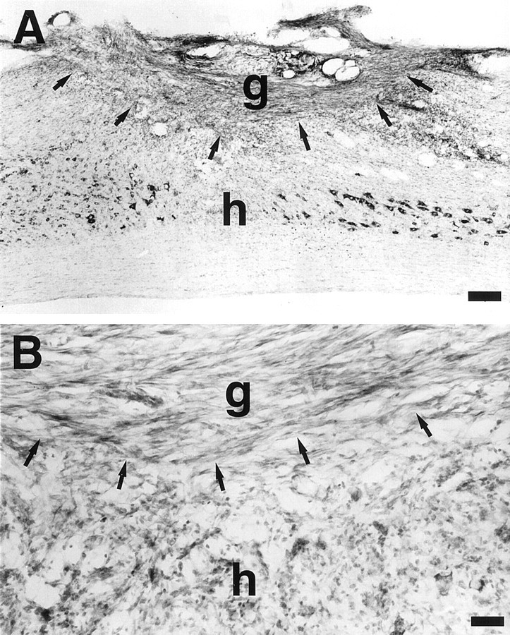
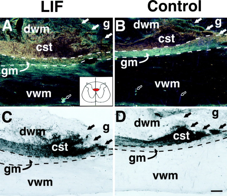
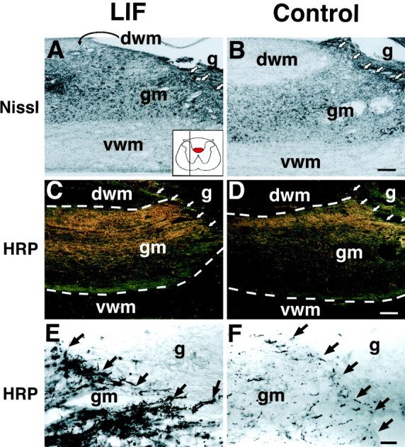
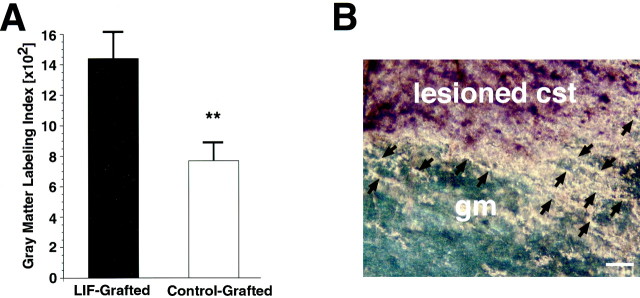
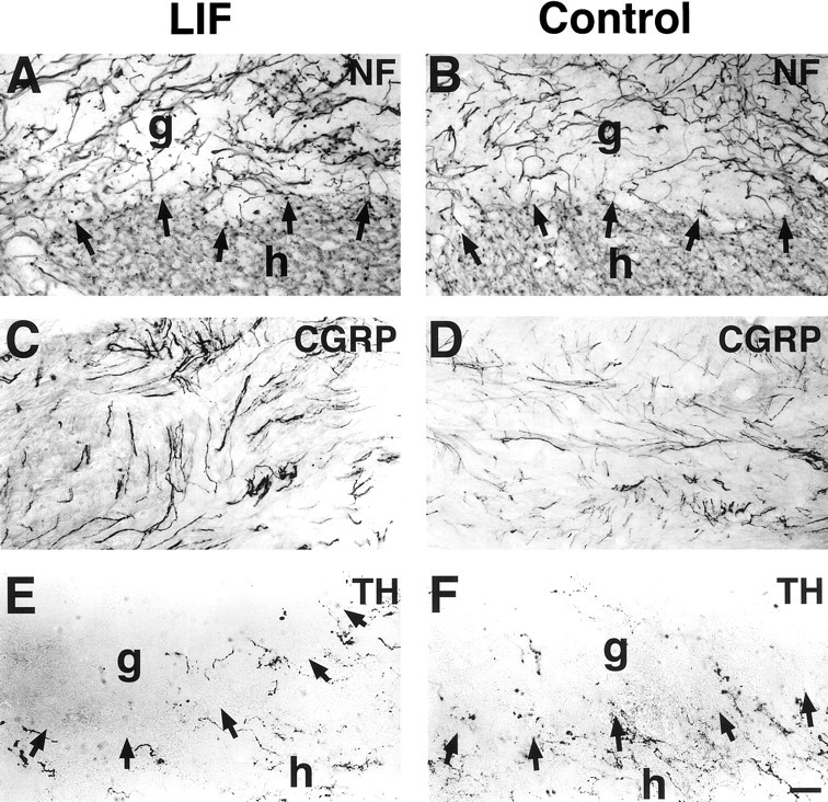
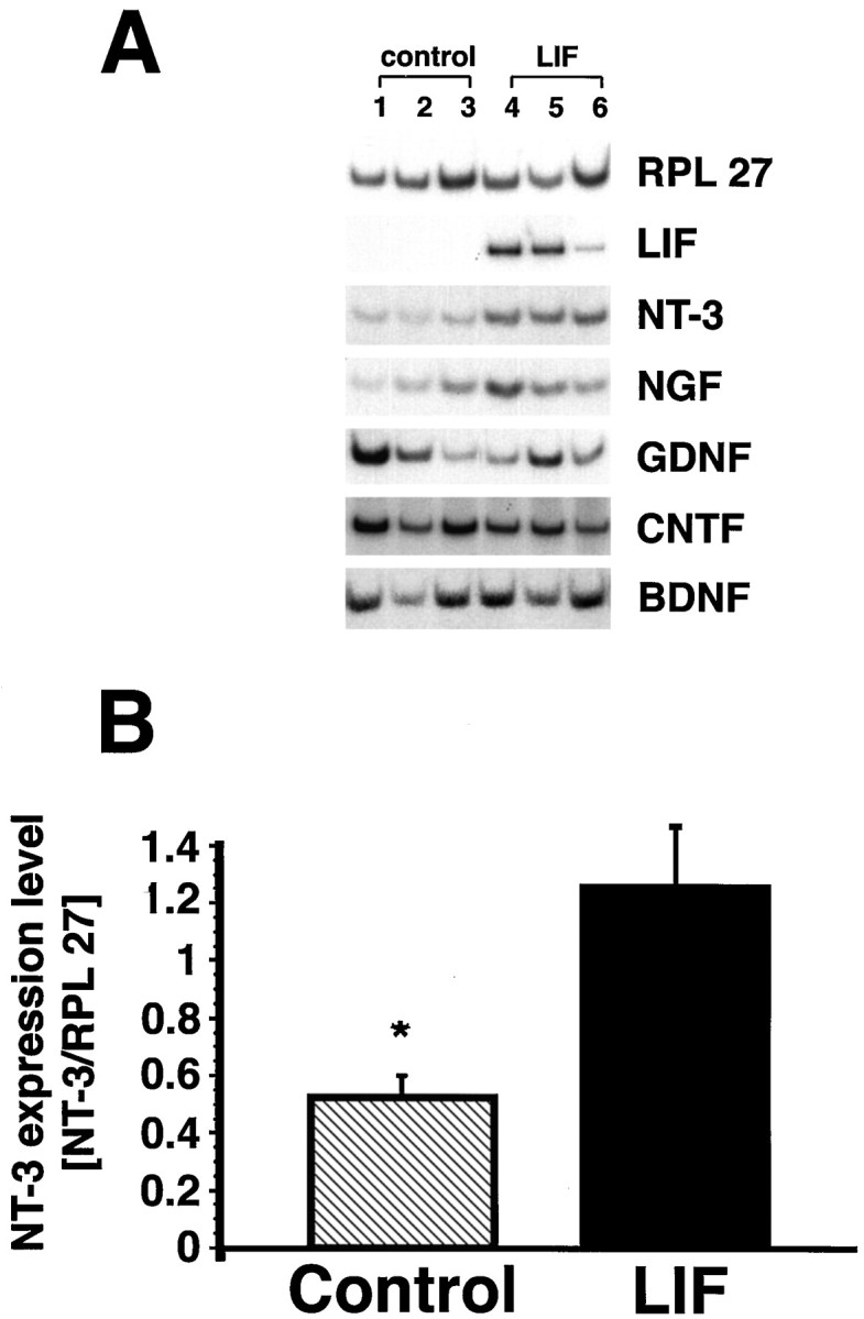
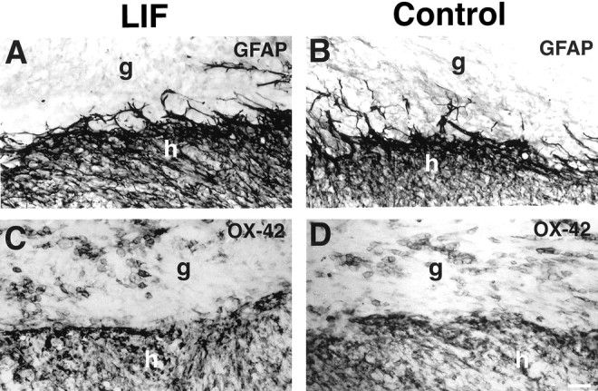
The cytokine leukemia inhibitory factor (LIF) modulates glial and neuronal function in development and after peripheral nerve injury, but little is known regarding its role in the injured adult CNS. To further understand the biological role of LIF and its potential mechanisms of action after CNS injury, effects of cellularly delivered LIF on axonal growth, glial activation, and expression of trophic factors were examined after adult mammalian spinal cord injury. Fibroblasts genetically modified to produce high amounts of LIF were grafted to the injured spinal cords of adult Fischer 344 rats. Two weeks after injury, animals with LIF-secreting cells showed a specific and significant increase in corticospinal axon growth compared with control animals. Furthermore, expression of neurotrophin-3, but not nerve growth factor, brain-derived neurotrophic factor, glia cell line-derived neurotrophic factor, or ciliary neurotrophic factor, was increased at the lesion site in LIF-grafted but not in control subjects. No differences in astroglial and microglial/macrophage activation were observed. Thus, LIF can directly or indirectly modulate molecular and cellular responses of the adult CNS to injury. These findings also demonstrate that neurotrophic molecules can augment expression of other trophic factors in vivo after traumatic injury in the adult CNS.


Figures

Fig. 1.
Fig. 1.
Characterization of LIF-expressing fibroblasts.A, Northern blot showing LIF RNA expression in LIF-transfected but not in control-transfected cells.LIF, LIF- expressing cells; β-gal, β-galactosidase-expressing cells. B, Western blot. A band of 45 kDa is visible in LIF-transfected (arrow) but not in control-transfected fibroblasts. The molecular weight of the marker proteins is indicated in lane 3.C, Growth of TF-1 cells is significantly higher when cocultured with LIF-transfected fibroblasts than with control β-gal-expressing cells. p < 0.01; mean ± SEM.
Fig. 2.
Fig. 2.
Integration of grafted fibroblasts into the host spinal cord. (A) Low- and (B) high-magnification view of Nissl-stained sagittal sections show numerous surviving grafted fibroblasts 2 weeks after grafting that span the dorsal hemisection lesion cavity (arrows) and provide a potential tissue bridge for growing axons. There is excellent graft integration into the lesion site. Arrows indicate host/graft interface. An LIF-secreting cell graft is pictured. g, Graft;h, host. Scale bars: A, 230 μm;B, 45 μm.
Fig. 3.
Fig. 3.
WGA-HRP labeling of the main corticospinal tract. Dark-field (A, B) and light-field (C, D) illumination show that the labeling intensity of the main corticospinal tract (cst) rostral to the injury site is similar in LIF-grafted (A, C) and control-grafted (B, D) animals. There is no significant retraction of CST axons from the lesion site at this time point (2 weeks after injury and grafting). Inset in A shows the approximate mediolateral location of the sections, indicating a relatively thin strip of central gray matter and wide strips of dorsal and ventral white matter; the red portion ofinset indicates the position of the dorsal CST. Scale bar: A, B, 172 μm; C, D, 220 μm. Host/graft interface is outlined by straight, filled arrows; the border between dorsal white matter (dwm), gray matter (gm), and ventral white matter (vwm) is indicated by dashed lines. g, Graft. Open arrows inA and B indicate the type of artifacts that were edited from digitized images before quantification of WGA-HRP grain density (see Materials and Methods).
Fig. 4.
Fig. 4.
Enhanced growth of lesioned corticospinal tract axons in LIF-grafted animals. Nissl stains (A, B) show the relative amounts of host gray matter and white matter in sections close to those pictured in C–F. A, LIF-grafted subject; B, control-grafted subject.dwm, Dorsal white matter; gm, gray matter; vwm, ventral white matter. Arrowsindicate host/graft (g) interface.Inset in A demonstrates that these sections are sampled from regions lateral to the midline; thus, dorsal white matter containing the CST (red ininset) is no longer visible in these sections. WGA-HRP-labeled sections (C–F) demonstrate substantially augmented growth of WGA-HRP-labeled CST axons up to the host/graft interface in host gray matter ventral to the lesion site in LIF-grafted (C, E) but not in control-grafted (D, F) animals. The preponderance of this growth is located in the more dorsally located gray matter rather than in ventrally located gray matter. In dark-field illumination (C, D), the borders between dorsal white matter (dvm), gray matter (gm), and ventral white matter (vwm) can be readily distinguished (indicated bydashed lines). Linear arrays of CST axons of the sort present in the gray matter of these lesioned spinal cords (E, F) are detectable only after injury; normally, CST-labeled ramifications in the gray matter exhibit numerous arborizations of fine, varicose axons but not linear growth. Scale bars: A, B, 220 μm; C, D, 172 μm;E, F, 42 μm.
Fig. 5.
Fig. 5.
A, Quantification of corticospinal WGA-HRP-labeled processes in spinal cord gray matter. At the lesion site, there is a significant increase in the number of WGA-HRP-labeled axonal processes in LIF-grafted recipients (p < 0.01; mean ± SEM). At this relatively brief time point, growth is not increased in an adjacent field 0.8 mm distal to the lesion. Labeling was quantified as described in Materials and Methods. B, Extending corticospinal tract axons originate from the lesioned dorsal CST. Dark-field photomicrograph from LIF-grafted subject demonstrates WGA-HRP-labeled axons extending linear profiles from the dorsal lesioned corticospinal tract (lesioned cst) into the underlying gray matter (gm) at the lesion site. Arrowsindicate individual axons. Scale bar, 34 μm.
Fig. 6.
Fig. 6.
Responses of other axonal systems to LIF-secreting and control grafts. Augmentation in the growth of other axonal systems is not observed in LIF-grafted recipients, indicating specificity of the growth effect to corticospinal systems. Neurofilament labeling (NF) shows no difference in overall axonal penetration into LIF-secreting (A) and control (B) grafts. Neurites that do penetrate grafts are mainly of sensory origin and exhibit immunolabeling for CGRP (C, LIF-secreting graft; D, control graft). There is also no difference in penetration ofTH-labeled coerulospinal axons adjacent to and within grafts (E, LIF-secreting; F, control grafts). Similarly, serotonergic and cholinergic axons penetrate grafts to a modest extent that does not significantly differ between graft types (data not shown). Arrows indicate host (h)/graft (g) interface. Scale bar: A, B, 55 μm; C–F, 42 μm.
Fig. 7.
Fig. 7.
LIF-secreting cells augment NT-3 gene expressionin vivo. A, Semiquantitative RT-PCR demonstrates clearly upregulated NT-3 expression in all LIF-grafted animals compared with control-grafted animals. No differences in NGF, BDNF, CNTF, and GDNF expression exist between LIF and control grafts (NGF expression is slightly but not significantly higher). Continued LIF transgene expression is evident in all LIF-grafted animals through the experimental period (2 weeks). B, Measurement of neurotrophin signal intensity shows significantly higher expression of NT-3 in LIF-grafted compared with control-grafted animals (p < 0.05 for NT-3; mean ± SEM). Units for NT-3 expression levels are provided as the ratio of NT-3 to RPL 27 expression, as a means of controlling for small differences in RNA amounts that may be present.
Fig. 8.
Fig. 8.
Microglial and astroglial activation at the host/graft interface. A, B, GFAP-immunolabeled processes are present at the host (h)/graft (g) interface, but extend relatively few processes into the graft. Qualitative and quantitative differences in GFAP labeling are not evident when comparing LIF-secreting (A) and control (B) grafts. Macrophages/microglia surround and invade (C) LIF-secreting and (D) control grafts to a similar extent, as shown by OX-42 labeling. Scale bar, 55 μm.

References

    1. Aloisi F, Rosa S, Testa U, Bonsi P, Russo G, Peschle C, Levi G. Regulation of leukemia inhibitory factor synthesis in cultured human astrocytes. J Immunol. 1994;152:5022–5031. - PubMed
    1. Apfel SC, Wright DE, Wiideman AM, Dormia C, Snider WD, Kessler JA. Nerve growth factor regulates the expression of brain-derived neurotrophic factor mRNA in the peripheral nervous system. Mol Cell Neurosci. 1996;7:134–142. - PubMed
    1. Arakawa Y, Sendtner M, Thoenen H. Survival effect of ciliary neurotrophic factor (CNTF) on chick embryonic motoneurons in culture: comparison with other neurotrophic factors and cytokines. J Neurosci. 1990;10:3507–3515. - PMC - PubMed
    1. Banner LR, Patterson PH. Major changes in the expression of the mRNAs for cholinergic differentiation factor/leukemia inhibitory factor and its receptor after injury to adult peripheral nerves and ganglia. Proc Natl Acad Sci USA. 1994;91:7109–7113. - PMC - PubMed
    1. Banner LR, Moayeri NN, Patterson PH. Leukemia inhibitory factor is expressed in astrocytes following cortical brain injury. Exp Neurol. 1997;147:1–9. - PubMed

Publication types

MeSH terms