. 1999 Apr 15;13(8):946-53.
doi: 10.1101/gad.13.8.946.

The subcellular localization of PBX1 and EXD proteins depends on nuclear import and export signals and is modulated by association with PREP1 and HTH

Affiliations

The subcellular localization of PBX1 and EXD proteins depends on nuclear import and export signals and is modulated by association with PREP1 and HTH

J Berthelsen et al. Genes Dev. .

Abstract

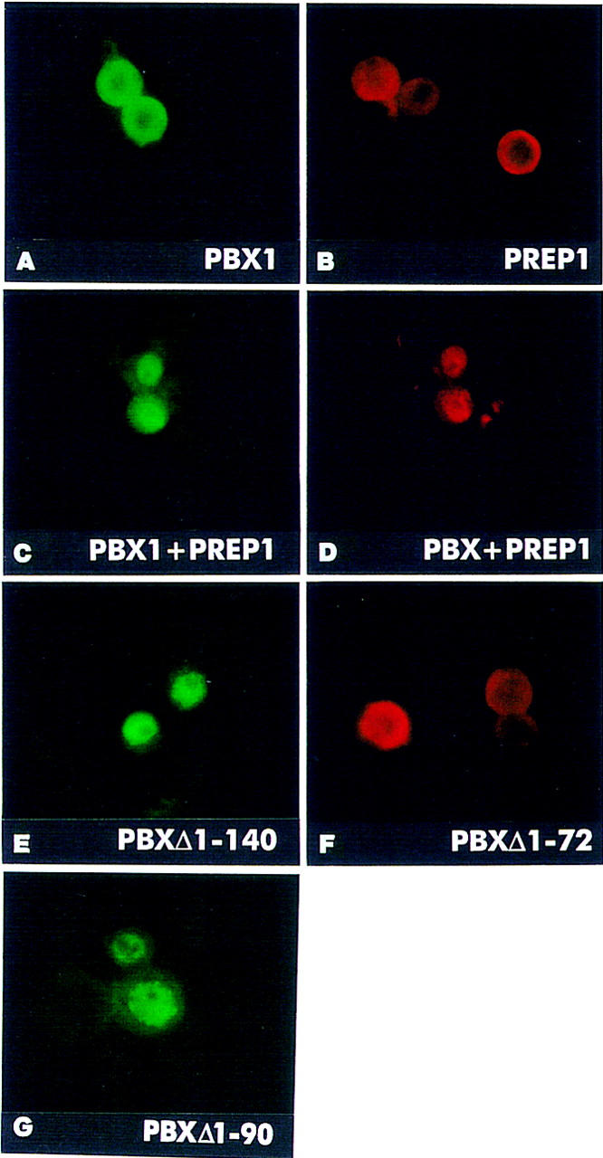
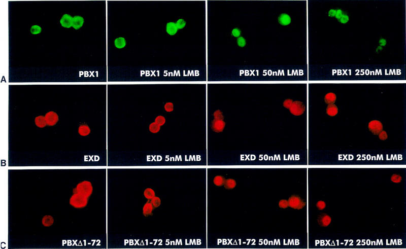
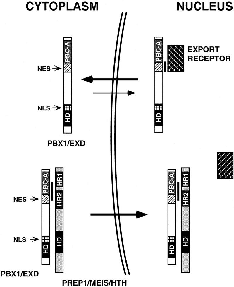
Nuclear localization of the Extradenticle (EXD) and PBX1 proteins is regionally restricted during Drosophila and mammalian development. We studied the subcellular localization of EXD, PBX, and their partners Homothorax (HTH) and PREP1, in different cell contexts. HTH and PREP1 are cytoplasmic and require association with EXD/PBX for nuclear localization. EXD and PBX1 are nuclear in murine fibroblasts but not in Drosophila Schneider cells, in which they are actively exported to the cytoplasm. Coexpression of EXD/PBX with HTH/PREP1 causes nuclear localization of their heterodimers in both cell contexts. We propose that heterodimerization with HTH/PREP induces nuclear translocation of EXD and PBX1 in specific cell contexts by blocking their nuclear export.


Figures

Figure 1
Figure 1
PREP1 is found in the cytoplasm of transfected mouse fibroblast cells and requires interaction with PBX1, which possesses a NLS within its homeodomain, for nuclear translocation. NIH-3T3 cells were transiently transfected with expression constructs for the indicated proteins and processed for indirect immunofluorescence with anti-PBX1 or anti-PREP1 polyclonal antibodies. PBX1 is nuclear (A), whereas PREP1 (B) is found in the cytoplasm of expressing NIH-3T3 cells. Cells coexpressing PREP1 and PBX1 (in red and green, respectively) display nuclear localization of both proteins (C,D). The PBXΔ1–140 mutant is nuclear in expressing cells (E,G, green), but is unable to trigger nuclear translocation of PREP1 (F, red). A PBX1 mutant lacking the homeodomain, PBXNT, is cytoplasmic in expressing NIH-3T3 cells (H, green). When coexpressed, PREP1 (red) and PBXNT (green) are both found in the cytoplasm of NIH-3T3 cells (I and J, respectively). Fusions of PREP1 with the SV40 NLS (NLS–PREP1), or with the PBX1 homeodomain (PREP/PBXHD) are nuclear (K,L, red). The reciprocal chimeric protein PBX/PREPHD is found in the cytoplasm (M, green). Double stainings (C,D,I,J) were performed with anti-HA rat monoclonal, to detect HA-tagged PBX1 and PBXNT, and anti-PREP1 polyclonal antibodies.
Figure 2
Figure 2
The EXD/PBX1 and HTH/PREP1 proteins are mutually required for nuclear localization in transfected Drosophila Schneider cells. A subregion of the PBC-A domain is necessary for cytoplasmic localization of PBX1 in Schneider cells. Drosophila Schneider cells were transiently transfected with expression constructs for the indicated proteins, and processed for indirect immunofluorescence with anti-PREP1 and anti-PBX1 polyclonal, or anti-FLAG and anti-HA monoclonal antibodies. PBX1 (A, green) and PREP1 (B, red) are found in the cytoplasm of expressing Schneider cells. Coexpression of PREP1 with PBX1 (C, green) causes nuclear localization of PREP1 (D, red). A PBX1 mutant, representing a deletion of its amino-terminal PBC-A domain, PBXΔ1–140 (E, green), is nuclear in Schneider cells in the absence of PREP1 or HTH. In contrast, the PBXΔ1–72 mutant (F, red) is found in the cytoplasm. PBXΔ1–90 is nuclear in expressing cells (G, green). Double stainings (C,D) were performed with anti-HA (rat) monoclonal, and anti-PREP1 polyclonal antibodies.
Figure 3
Figure 3
LMB blocks nuclear export of PBX1, EXD, and PBXΔ1–72 proteins in Drosophila Schneider cells. Cells were transiently transfected with expression constructs for PBX1, EXD, and PBXΔ1–72 and processed for indirect immunofluorescence with anti-PBX1 and anti-Flag antibodies. Schneider cells, transfected with the PBX1, the EXD or the PBXΔ1–72 expressors were treated with 5, 50, and 250 nm LMB as indicated. Fifty and 250 nm LMB induce relocalization of PBX1 (A, green), of EXD (B, red), or of the PBXΔ1–72 mutant (C, red) into the nuclei of expressing cells. Treatment with LMB at the indicated concentrations was performed 18 hr after transfection.
Figure 4
Figure 4
A model for the regulation of subcellular localization of PBC and PREP1/MEIS/HTH proteins in cells displaying cytoplasmic localization of PBC proteins. PBC and PREP1/MEIS/HTH proteins are represented schematically. In specific cell contexts (e.g., Schneider cells), in the absence of PREP1/MEIS/HTH proteins, PBC proteins are actively exported from the nucleus, a process requiring sequences (NES, hatched box) located within their conserved PBC-A domain, which are recognized by a nuclear export receptor (dark, squared rectangle). PBC proteins form stable complexes with PREP1/MEIS/HTH proteins, when coexpressed, through an interaction surface that coincides with the region required for nuclear export, thereby shielding it. The newly formed complex translocates into the nucleus owing to the NLS located within the homeodomain of PBC proteins (NLS, white-squared box). Black rectangles represent the homeodomains (HD). Light gray and dark gray boxes represent conserved amino-terminal regions within PBC and PREP1/MEIS/HTH proteins, respectively (PBC-A, HR1/HR2). A black vertical line indicates protein–protein contacts.

References

    1. Abu-Shaar M, Mann R. Generation of multiple antagonistic domains along the proximodistal axis during Drosophila leg development. Development. 1998;125:3821–3830. - PubMed
    1. Berthelsen J, Zappavigna V, Ferretti E, Mavilio F, Blasi F. The novel homeoprotein Prep1 modulates Pbx-Hox protein cooperativity. EMBO J. 1998a;17:1434–1445. - PMC - PubMed
    1. Berthelsen J, Zappavigna V, Mavilio F, Blasi F. Prep1, a novel functional partner of Pbx proteins. EMBO J. 1998b;17:1423–1433. - PMC - PubMed
    1. Burglin T. Analysis of TALE superclass homeobox genes (MEIS, PBC, KNOX, Iroquois, TGIF) reveals a novel domain conserved between plants and animals. Nucleic Acids Res. 1997;25:4173–4180. - PMC - PubMed
    1. Burglin TR, Ruvkun G. New motif in PBX genes. Nat Genet. 1992;1:319–320. - PubMed

Publication types

Grants and funding

LinkOut - more resources