. 1999 Apr 15;13(8):954-65.
doi: 10.1101/gad.13.8.954.

ebi regulates epidermal growth factor receptor signaling pathways in Drosophila

Affiliations

ebi regulates epidermal growth factor receptor signaling pathways in Drosophila

X Dong et al. Genes Dev. .

Abstract

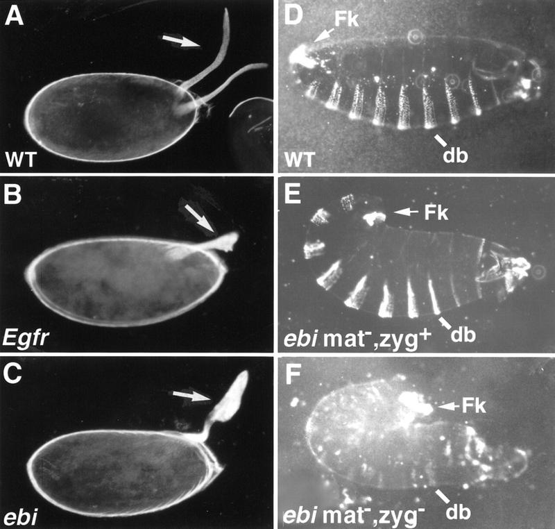
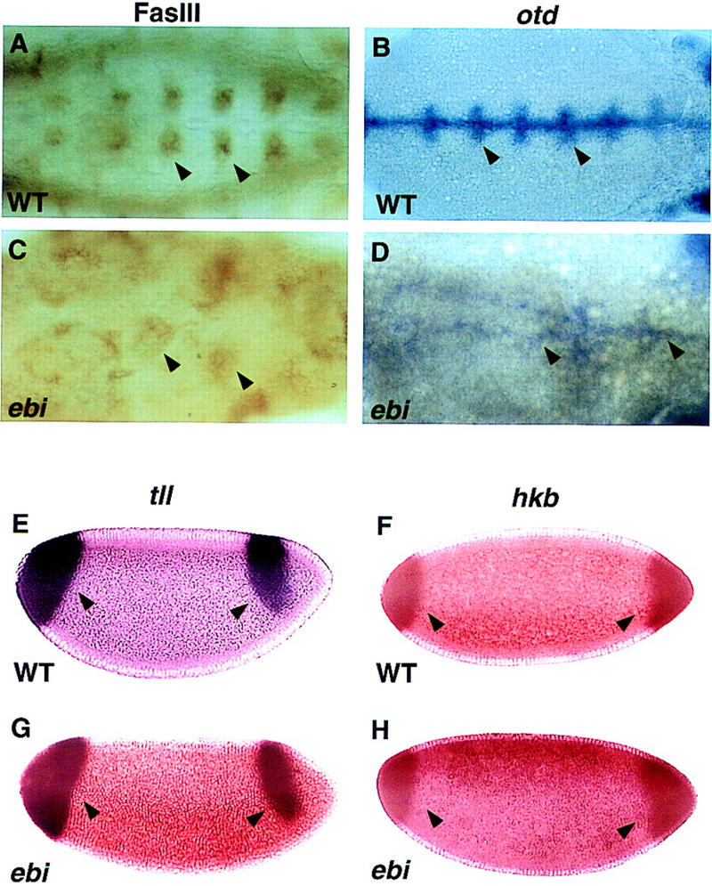
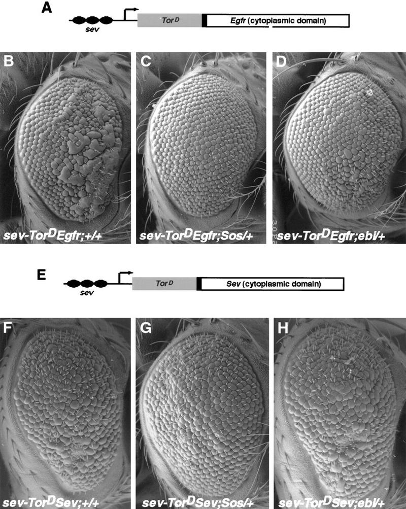
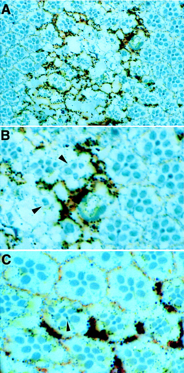
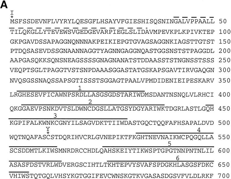
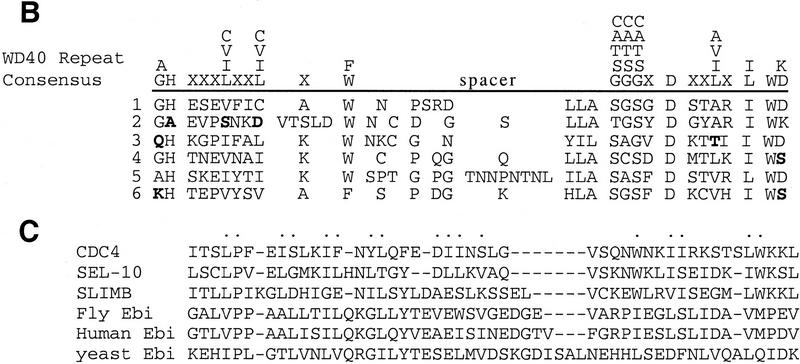
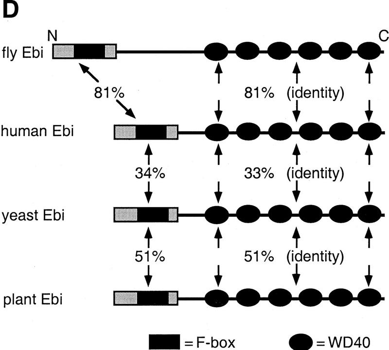
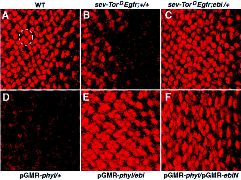
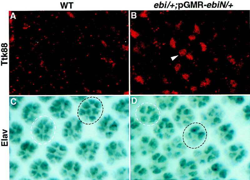
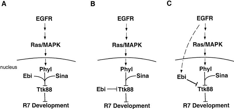
ebi regulates the epidermal growth factor receptor (EGFR) signaling pathway at multiple steps in Drosophila development. Mutations in ebi and Egfr lead to similar phenotypes and show genetic interactions. However, ebi does not show genetic interactions with other RTKs (e.g., torso) or with components of the canonical Ras/MAP kinase pathway. ebi encodes an evolutionarily conserved protein with a unique amino terminus, distantly related to F-box sequences, and six tandemly arranged carboxy-terminal WD40 repeats. The existence of closely related proteins in yeast, plants, and humans suggests that ebi functions in a highly conserved biochemical pathway. Proteins with related structures regulate protein degradation. Similarly, in the developing eye, ebi promotes EGFR-dependent down-regulation of Tramtrack88, an antagonist of neuronal development.


Figures

Figure 1
Figure 1
ebi mutants exhibit egg and embryonic cuticle phenotypes. (A) Two dorsal appendages (arrow) are located at the dorsal anterior region of the wild-type egg. (B) Females harboring a weak Egfr allele, Egfrtop1/Egfrtop1, produce ventralized eggs with fused dorsal appendages (arrow). (C) Eggs laid by females mutant for ebi (ebiE90/ebiP7) are also ventralized (arrow). (D) Wild-type embryos. (E) Embryos lacking maternal, but not zygotic, contribution of ebi exhibit a tail-up embryo, indicating failure of germ-band retraction. These embryos also have head defects. Embryos lacking maternal ebi were generated using the ovoD method (see Materials and Methods). (F) Embryos lacking maternal and zygotic ebi exhibit more severe phenotypes, including loss of ventral denticle belt (db) structures and a tightly curled morphology. In addition, severe head defects and malformed posterior filzkörper (Fk) were observed. Anterior is to the right.
Figure 2
Figure 2
ebi is required for EGFR but not Torso receptor-dependent gene expression. RTK-dependent gene expression in wild-type embryos (A,B,E,F) and embryos lacking both maternal and zygotic ebi (C,D,G,H) were compared. The expression of otd and FasIII is EGFR dependent. Expression of tll and hkb is Torso dependent. (A,C) FasIII expression was determined by immunohistology using mAb7G10. A ventral view of stage 11/12 embryos is shown. (A) In wild-type embryos, FasIII is expressed in clusters of cells flanking the midline of the ventral ectoderm (arrowheads). (C) FasIII levels are severely reduced in ebi embryos (arrowheads). (B,D) Expression of otd mRNA was detected by in situ hybridization. A ventral view of stage 10/11 embryos is shown. (B) Wild-type embryos show strong otd staining in ventral-most ectoderm (arrowheads). (D) Although otd expression is markedly reduced in ebi mutant embryos, residual staining is observed (arrowheads). Expression of tll and hkb mRNA in ebi (G,H) is indistinguishable from wild-type (E,F, respectively). Anterior is to the right.
Figure 3
Figure 3
ebi is a dominant suppressor of ectopic R7 development induced by an activated EGFR. (A) A schematic representation of the sev–TorDEgfr construct (Reichman-Fried et al. 1994). (B–D) Scanning electron micrographs of adult eyes showing sev–TorDEgfr alone (B), in an SosSF15/+ background (C), and in an ebiE90/+ background. Like Sos, ebi is a dominant suppressor of sev–TorDEgfr. (E) A schematic representation of sev–TorDSev (Dickson et al. 1992b). (F–H) Scanning electron micrographs of adult eyes showing sev-TorDSev alone (F), in an SosSF15/+ background (G) and in an ebiE90/+ background (H). Although Sos suppresses the sev–TorDSev phenotype, ebi does not (see Table 1). Anterior is to the right.
Figure 4
Figure 4
ebi ommatidia are highly abnormal. ebi homozygous clones were generated using FRT-mediated mitotic recombination catalyzed by Flp recombinase expressed under the control of the ey promoter. Pigmented regions are homozygous for the ebiE4 allele; pigmentation results from two copies of a w+ transgene on the ebiE4 chromosome (see Materials and Methods). Ommatidia heterozygous for w+ transgene or lacking this transgene are lightly pigmented or appear unpigmented in the sections. (A) Wild-type ommatidia in the unpigmented region are separated by a pigmented region containing highly abnormal ommatidia. (B) Many ommatidia within the labeled regions lack R cells (see arrowheads). (C) Although many mutant ommatidia lack R cells of all different classes, some normal looking ommatidia contain both ebi mutant and wild-type cells. The arrow indicates an ommatidium containing a genotypically mutant R7 cell (i.e., containing pigment granules).
Figure 5
Figure 5
ebi encodes a protein containing WD40 repeats and a diverged F box. (A) The amino acid sequence of Ebi. The F-box-like sequence is indicated by the broken line. WD40 repeats are indicated by solid lines. Missense mutations in ebiE4 (amino acid 1) and ebiE90 (amino acid 510) alleles are indicated above the sequence. (B) Alignment of WD40 repeats. The WD40 consensus sequence according to Neer et al. (1994) is shown. Divergence from the consensus in the Ebi repeats are indicated in boldface type. (C) Alignment of F-box sequence with Ebi amino-terminal sequence (see text). Dots above the alignment indicate the conserved residues in F boxes according to Patton et al. (1998) (D) Schematic representation of fly, human, yeast, and plant Ebi proteins. The percentage of identical amino acids in each region relative to fly Ebi is indicated (see text).
Figure 5
Figure 5
ebi encodes a protein containing WD40 repeats and a diverged F box. (A) The amino acid sequence of Ebi. The F-box-like sequence is indicated by the broken line. WD40 repeats are indicated by solid lines. Missense mutations in ebiE4 (amino acid 1) and ebiE90 (amino acid 510) alleles are indicated above the sequence. (B) Alignment of WD40 repeats. The WD40 consensus sequence according to Neer et al. (1994) is shown. Divergence from the consensus in the Ebi repeats are indicated in boldface type. (C) Alignment of F-box sequence with Ebi amino-terminal sequence (see text). Dots above the alignment indicate the conserved residues in F boxes according to Patton et al. (1998) (D) Schematic representation of fly, human, yeast, and plant Ebi proteins. The percentage of identical amino acids in each region relative to fly Ebi is indicated (see text).
Figure 6
Figure 6
Ebi is a widely expressed nuclear protein. Third-instar eye discs and embryos were stained with an affinity purified anti-Ebi antibody (see Materials and Methods). (A) Ebi is expressed in the nuclei of all cells in the eye disc. (MF) Morphogenetic furrow. Nuclei of cells in the morphogenetic furrow are out of the focal plane shown. High magnification views of a five-cell precluster (B) and eight-cell cluster (C) are shown. The R7 precursor cell is indicated by the arrowhead in C. Anterior is to the right. (D) Ebi is widely expressed in nuclei (inset) of a stage 8 embryo, as well as throughout embryogenesis. (E) Salivary glands were stained with both anti-Myc antibodies to detect Myc-tagged Ebi (red) (see text and Materials and Methods) and the DNA stain DAPI (green). Ebi was not associated with chromosomes or the nucleolus (*) but, rather, was distributed in a reticular network pattern throughout the nucleoplasm.
Figure 7
Figure 7
ebi promotes Ttk88 protein degradation. Eye discs labeled with anti-Ttk88 (A–F). (A) In wild-type, Ttk88 is expressed in cone cell nuclei. A single ommatidium is circled to show four labeled cone cells. (B) Ttk88 staining in cone cells is severely reduced in eye discs expressing sev–TorDEgfr. Residual labeling was seen in the posterior region of eye discs, which correlates with the less severe phenotype seen in the posterior part of the adult eye (see Fig. 5B). (C) Ttk88 levels are restored in eye discs heterozygous for ebiE90. pGMR–phyl also induces Ttk88 degradation (D). (E) ebiE4 dominantly suppresses phyl-induced Ttk88 degradation. (F) pGMR-driven expression of a dominant negative ebi transgene, pGMR–ebiN, also restores Ttk88 protein levels in discs expressing pGMR–phyl.
Figure 8
Figure 8
Ttk88 is up-regulated in ebiP25/+; pGMR–ebiN/+ eye discs. (A,B) Anti-Ttk88 staining; (C,D) anti-Elav staining. (A) In wild type, Ttk88 is not expressed in a focal plane containing differentiating R cell nuclei. (B) In ebiP25/+; pGMR–ebiN/+ eye discs, Ttk88 is up-regulated in this region. (C) Wild-type eye disc. An eight-cell cluster, 11 rows posterior to the morphogenetic furrow, is encircled by a broken black line. The R7 cell is slightly out of the focal plane and appears only lightly stained. A more mature cluster, three rows posterior, is encircled by a broken white line. (D) ebiP25/+; pGMR–ebiN/+ eye disc. Two ommatidia containing less than eight Elav-stained R cells at equivalent stages of development as those shown in C are encircled.
Figure 9
Figure 9
Models for Ebi function in R7 induction. Data presented in this paper are consistent with the following models for Ebi function in R7 development. (A) Ebi functions in a common biochemical pathway with Phyl and Sina to down-regulate Ttk88; (B) Ebi is a signal-independent constitutive negative regulator of Ttk88; (C) Ebi participates in a parallel pathway regulating Ttk88 downregulation activated by the EGFR.

Similar articles

Cited by

References

    1. Altschul SF, Gish W, Miller W, Myers EW, Lipman DJ. Basic local alignment search tool. J Mol Biol. 1990;215:403–410. - PubMed
    1. Bai C, Sen P, Hofman K, Ma L, Goebl M, Harper JW, Elledge S. Skp1 connects cell cycle regulators to the ubiquitin proteolysis machinery through a novel motif, the F-box. Cell. 1996;86:263–274. - PubMed
    1. Banerjee U, Renfranz PJ, Pollock JA, Benzer S. Molecular characterization and expression of sevenless, a gene involved in neuronal pattern formation in the Drosophila eye. Cell. 1987;49:281–291. - PubMed
    1. Basler K, Christen B, Hafen E. Ligand-independent activation of the sevenless Receptor tyrosine kinase changes the fate of cells in the developing Drosophila eye. Cell. 1991;64:1069–1081. - PubMed
    1. Bronner G, Chu-LaGraff Q, Doe CQ, Cohen B, Weigel D, Taubert H, Jäckle H. Sp1/egr-like zinc-finger protein required for endoderm specification and germ-layer formation in Drosophila. Nature. 1994;369:664–668. - PubMed

Publication types

MeSH terms

Associated data