. 1999 Apr 15;13(8):978-86.
doi: 10.1101/gad.13.8.978.

Schwann cell hyperplasia and tumors in transgenic mice expressing a naturally occurring mutant NF2 protein

Affiliations

Schwann cell hyperplasia and tumors in transgenic mice expressing a naturally occurring mutant NF2 protein

M Giovannini et al. Genes Dev. .

Abstract

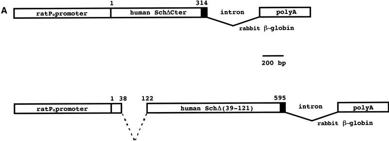
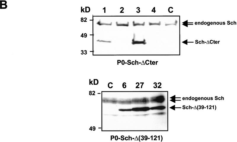
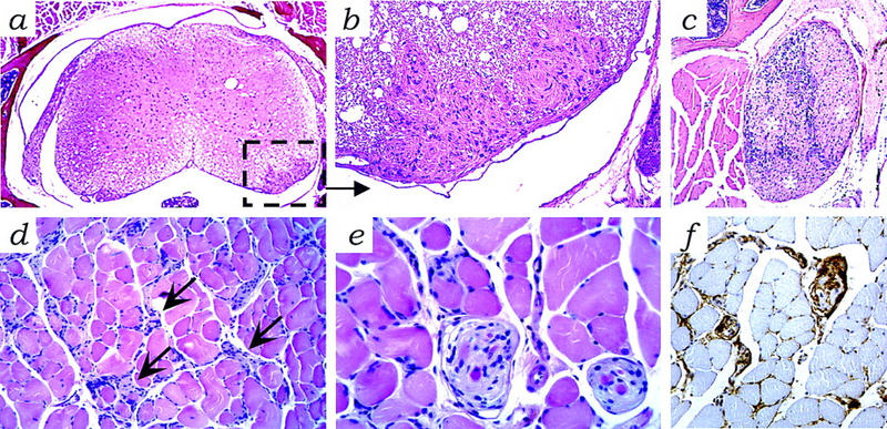
Specific mutations in some tumor suppressor genes such as p53 can act in a dominant fashion. We tested whether this mechanism may also apply for the neurofibromatosis type-2 gene (NF2) which, when mutated, leads to schwannoma development. Transgenic mice were generated that express, in Schwann cells, mutant NF2 proteins prototypic of natural mutants observed in humans. Mice expressing a NF2 protein with an interstitial deletion in the amino-terminal domain showed high prevalence of Schwann cell-derived tumors and Schwann cell hyperplasia, whereas those expressing a carboxy-terminally truncated protein were normal. Our results indicate that a subset of mutant NF2 alleles observed in patients may encode products with dominant properties when overexpressed in specific cell lineages.


Figures

Figure 1
Figure 1
Structure and expression of the transgenes. (A) Schematic representation of the P0–Sch-ΔCter and P0–Sch-Δ(39–121) transgenes. The transgenic constructs were made by inserting the VSV-tagged (black box) mutated human NF2 cDNAs into plasmid pPG6 between the 5′ flanking sequence from the rat P0 gene (1.1 kb) and the rabbit β-globin 3′ splicing plus polyadenylation signal (1.2 kb). (B) Transgene expression analysis by Western blotting. The 39-kD (Sch-ΔCter) and 62-kD Sch-Δ(39–121) schwannomin mutants were detected by immunoblotting with a polyclonal anti-NF2–Nter serum in protein extracts from sciatic nerve of the F1 mice of two of four P0–Sch-ΔCter and three of three P0–Sch-Δ(39–121) independent strains. The polyclonal anti-NF2–Nter A-19, raised against a peptide corresponding to amino acids 2–21 of human schwannomin, detects both the endogenous murine NF2 protein and the two transgenic mutant human schwannomins (arrows). (Lanes 1–4) Sciatic nerves from strains P0–Sch-ΔCter; (lanes 6,27,32) sciatic nerves from strains P0–Sch-Δ(39–121). (C) Sciatic nerves from nontransgenic FVB/N littermate. (C) To confirm that the mutant proteins are encoded by the transgene and to analyze the spatial profile of expression of the transgenes, total protein extracts were obtained from various organs of transgenic animals of strains P0–Sch-ΔCter/3 and P0–Sch-Δ(39–121)/6, 27, and 32 and analyzed by Western blotting. Transgenic proteins were readily detected by immunoblotting with anti-VSV polyclonal antibodies in peripheral nerves (trigeminal and sciatic nerves) and uterus. (SN) Sciatic nerve; (Br) brain; (Ce) cerebellum; (BS) brain stem; (TN) trigeminal nerve; (SC) spinal cord; (He) heart; (Ut) uterus; (Sp) spleen; (Lu) lung; (Ki) kidney; (Li) liver.
Figure 1
Figure 1
Structure and expression of the transgenes. (A) Schematic representation of the P0–Sch-ΔCter and P0–Sch-Δ(39–121) transgenes. The transgenic constructs were made by inserting the VSV-tagged (black box) mutated human NF2 cDNAs into plasmid pPG6 between the 5′ flanking sequence from the rat P0 gene (1.1 kb) and the rabbit β-globin 3′ splicing plus polyadenylation signal (1.2 kb). (B) Transgene expression analysis by Western blotting. The 39-kD (Sch-ΔCter) and 62-kD Sch-Δ(39–121) schwannomin mutants were detected by immunoblotting with a polyclonal anti-NF2–Nter serum in protein extracts from sciatic nerve of the F1 mice of two of four P0–Sch-ΔCter and three of three P0–Sch-Δ(39–121) independent strains. The polyclonal anti-NF2–Nter A-19, raised against a peptide corresponding to amino acids 2–21 of human schwannomin, detects both the endogenous murine NF2 protein and the two transgenic mutant human schwannomins (arrows). (Lanes 1–4) Sciatic nerves from strains P0–Sch-ΔCter; (lanes 6,27,32) sciatic nerves from strains P0–Sch-Δ(39–121). (C) Sciatic nerves from nontransgenic FVB/N littermate. (C) To confirm that the mutant proteins are encoded by the transgene and to analyze the spatial profile of expression of the transgenes, total protein extracts were obtained from various organs of transgenic animals of strains P0–Sch-ΔCter/3 and P0–Sch-Δ(39–121)/6, 27, and 32 and analyzed by Western blotting. Transgenic proteins were readily detected by immunoblotting with anti-VSV polyclonal antibodies in peripheral nerves (trigeminal and sciatic nerves) and uterus. (SN) Sciatic nerve; (Br) brain; (Ce) cerebellum; (BS) brain stem; (TN) trigeminal nerve; (SC) spinal cord; (He) heart; (Ut) uterus; (Sp) spleen; (Lu) lung; (Ki) kidney; (Li) liver.
Figure 2
Figure 2
Schwann cell tumors in P0–Sch-Δ(39–121) transgenic mice. (a) Moderately proliferative and locally aggressive schwannoma (asterisk) originating from the ganglion semilunaris of the trigeminal nerve that had infiltrated peripherally the perineurium reaching the pituitary stalk (arrow) and invaded the surrounding soft tissues down to the region of the soft palate and the external ear region (9-month-old male, strain 32), hematoxylin and eosin stain. The tumor displayed moderate pleiomorphism, dense cellularity, and areas of S-100 (b) protein and LNGFR immunoreactivity (c). (d) Schwannoma arising from a paravertebral spinal ganglion in a 17-month-old mouse (strain 27) that presented a generalized increase in Schwann cells in various ganglia. The tumor was composed of loosely arranged interwoven bundles of fusiform cells within scant fibrovascular stroma. Tumor cells had indistinct borders and the cytoplasm blended into the stroma, hematoxylin and eosin. (e) Representative microscopic features of a uterine tumor. Compactly arranged spindle-shaped cells in tumor 4228 (17-month-old female; strain 6), hematoxylin and eosin stain. (f) Focal areas of weak S-100 protein and (g) strong, diffuse LNGFR immunoreactivity in uterine tumor 4235 (20-month-old female; strain 27). (h) Schwann cell proliferation at the internal surface of a Peyer’s plaque in a 20-month-old female (strain 27), hematoxylin and eosin stain, showing (j) weak S-100 protein, and (k) strong LNGFR immunoreactivity. Magnification, 25× (a); 200× (b,c); 100× (d); 400× (e–g); 100× (h–k).
Figure 3
Figure 3
P0–Sch-Δ(39–121) transgene expression in tumors. Immunoprecipitations were performed on RIPA extracts of the different tumors with polyclonal antibody anti-NF2–Cter C-18. Immunoprecipitated proteins were separated on a 8% polyacrylamide gel and electrotransferred to nitrocellulose membrane. Immunoblottings were performed with anti-NF2–Nter A-19 polyclonal antibody (top) and anti-VSV-G monoclonal antibody (bottom). Detection was performed by chemoluminescence and the exposure time was defined for optimal detection of transgene expression in uterine tumors (lanes 1–7). Under these conditions, transgene expression in the normal transgenic uterus (lane 9) is hardly detectable. (Lane 1) Uterine tumor (strain 6); (lanes 2–7) uterine tumors (strain 27); (lane 8), lung adenocarcinoma (strain 27); (lane 9) normal transgenic uterus (strain 32).
Figure 4
Figure 4
Tumors from P0–Sch-Δ(39–121) transgenic mice are diploid. FISH with mouse chromosome 11-specific probe on paraffin sections. The biotin-labeled probe was detected with FITC-avidin and nuclei were visualized by counterstaining with propidium iodide. Nuclei from a normal trigeminal nerve (a) and from a uterine tumor (b) in which expression of wild-type schwannomin was shown by immunoprecipitation analysis (same tumor as in Fig. 3, lane 1), were taken as reference. The presence of two distinct hybridization signals in the majority of the hybridized nuclei from both tumor and normal tissue proved that the tumor was diploid for chromosome 11. Nuclei showing two distinct hybridization signals (c) in a schwannoma originating from the trigeminal nerve of a transgenic P0–Sch-Δ(39–121) mouse (same tumor as in Fig. 2A), and in a schwannoma arising from a paravertebral spinal ganglion (d) (same tumor as in Fig. 2D). Magnification, 1250×.
Figure 5
Figure 5
Schwannosis in P0–Sch-Δ(39–121) transgenic tissues. (a) Microscopic foci of proliferated Schwann cells (schwannosis) in the spinal cord [inset; enlarged in b and c (asterisks)] in a spinal ganglion. (d) Hyperplastic Schwann cells and hypertrophic nerve bundles (arrows) in skeletal muscle with (e) focal aspects of nodular Schwann cell growth displaying (f) S-100 protein immunoreactivity. Schwannosis is a frequent histopathological feature of the peripheral nerves of NF2 patients (Wiestler and Radner 1994). Hematoxylin and eosin stain. Magnification, 25× (a); 200× (b,d,f); 100× (c); 400× (e).

References

    1. Akagi K, Sandig V, Vooijs M, Van der Valk M, Giovannini M, Strauss M, Berns A. Cre-mediated somatic site-specific recombination in mice. Nucleic Acids Res. 1997;25:1766–1773. - PMC - PubMed
    1. Bianchi AB, Hara T, Ramesh V, Gao J, Klein-Szanto AJ, Morin F, Menon AG, Trofatter JA, Gusella JF, Seizinger BR, Kley N. Mutations in transcript isoforms of the neurofibromatosis 2 gene in multiple human tumour types. Nat Genet. 1994;6:185–192. - PubMed
    1. Bijlsma EK, Merel P, Bosch DA, Westerveld A, Delattre O, Thomas G, Hulsebos TJ. Analysis of mutations in the SCH gene in schwannomas. Genes Chromosomes Cancer. 1994;11:7–14. - PubMed
    1. Deguen B, Merel P, Goutebroze L, Giovannini M, Reggio H, Arpin M, Thomas G. Impaired interaction of naturally occurring mutant NF2 protein with actin-based cytoskeleton and membrane. Hum Mol Genet. 1998;7:217–226. - PubMed
    1. Eldridge R. Central neurofibromatosis with bilateral acoustic neuroma. Adv Neurol. 1981;29:57–65. - PubMed

Publication types