. 1999 Apr 15;13(8):1015-24.
doi: 10.1101/gad.13.8.1015.

TRAF6 deficiency results in osteopetrosis and defective interleukin-1, CD40, and LPS signaling

Affiliations

TRAF6 deficiency results in osteopetrosis and defective interleukin-1, CD40, and LPS signaling

M A Lomaga et al. Genes Dev. .

Abstract

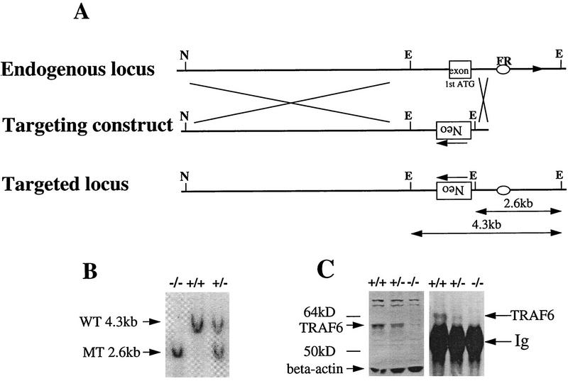
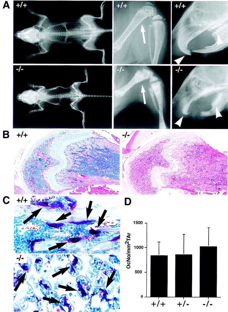
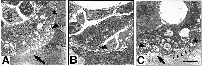
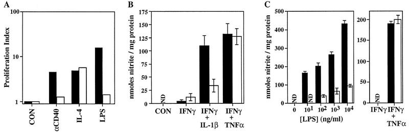
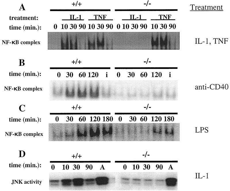
Bone resorption and remodeling is an intricately controlled, physiological process that requires the function of osteoclasts. The processes governing both the differentiation and activation of osteoclasts involve signals induced by osteoprotegerin ligand (OPGL), a member of tumor necrosis factor (TNF) superfamily, and its cognate receptor RANK. The molecular mechanisms of the intracellular signal transduction remain to be elucidated. Here we report that mice deficient in TNF receptor-associated factor 6 (TRAF6) are osteopetrotic with defects in bone remodeling and tooth eruption due to impaired osteoclast function. Using in vitro assays, we demonstrate that TRAF6 is crucial not only in IL-1 and CD40 signaling but also, surprisingly, in LPS signaling. Furthermore, like TRAF2 and TRAF3, TRAF6 is essential for perinatal and postnatal survival. These findings establish unexpectedly diverse and critical roles for TRAF6 in perinatal and postnatal survival, bone metabolism, LPS, and cytokine signaling.


Figures

Figure 1
Figure 1
Disruption of the murine traf6 gene by homologous recombination. (A). Restriction enzyme maps of the 5′ region of the endogenous traf6 locus (top) and the targeting construct (middle) are shown. The targeting vector was engineered to replace the exon containing the ATG translation initiation codon with PGK-neo in the opposite orientation (bottom). This exon encodes the first 100 amino acids of TRAF6, representing a portion of the amino-terminal ring-finger domain. EcoRI digestion generates a 4.3-kb wild-type band and a 2.6-kb mutant band. The flanking region (FR) probe used for Southern blot analysis is represented by an oval. (E) EcoRI; (N) NotI. (B) Southern blot analysis of genomic DNA from primary embryonic fibroblasts harvested from E14.5 wild-type (+/+), heterozygous (+/−) and homozygous (−/−) TRAF6 embryos. DNA was digested with EcoRI and hybridized with radiolabeled FR. Arrows indicate the 4.3-kb wild-type (WT) and the 2.6-kb mutant (MT) bands. (C) Immunoprecipitation and Western blot analysis of TRAF6 protein. TRAF6 was immunoprecipitated from total kidney lysate of TRAF6+/+, TRAF6+/−, and TRAF6−/− mice using polyclonal anti-TRAF6 antibody raised against the full-length TRAF6 protein. Western blots of both total lysates (left) and immunoprecipitates (right) were probed with the same antibody. The arrow indicates the presence of the 60-kD TRAF6 protein in +/+ lysates, a reduced level of TRAF6 in +/− lysates, and the absence of TRAF6 in −/− lysates.
Figure 2
Figure 2
Severe osteopetrosis in the presence of TRAP+ osteoclasts in TRAF6−/− mice. (A) X-ray analysis of the bone density of a 4-week-old TRAF6 knockout (−/−) and a wild-type littermate (+/+) mouse. A complete scan of the skeletons of both mice, including their femurs, tibia/fibulas, and facial regions was performed. Note the increase in overall bone density in the TRAF6−/− mouse, the shortening of its long bones, the enlargement of its metaphyses (arrow), as well as the absence of incisors and molars (arrowheads) in its oral cavity. Skeletal structure and morphogenesis of flat bones appears normal. (B) H&E staining depicting histological changes in bone. The shaft of the femur in TRAF6−/− mice is filled with cartilage and bone. There is some evidence of periosteal bone modeling occurring adjacent to the growth plates. Magnification, 4×. (C) TRAP staining. Presence of multinucleate TRAP+ osteoclasts (arrows) in TRAF6−/− mice. Most TRAF6−/− osteoclasts are withdrawn from the bone surface, whereas osteoclasts in wild type mice are attached to the bone. Magnification 60×. (D) The number of TRAP+ osteoclasts per mm2 tissue area in TRAF6−/−, TRAF6+/−, and TRAF6+/+ mice is comparable.
Figure 3
Figure 3
Electron microscopy of osteoclasts in TRAF6−/− and TRAF6+/− mice. (A) TRAF6+/− osteoclast seen in a resorption lacuna. The depicted cell exhibits an attachment zone (asterisk), cytoplasmic vacuolization, and ruffled border (arrowhead), features of a normally activated, mineral-resorbing (arrow) osteoclast. (B) This cell illustrates the typical osteoclast in TRAF6−/− mice. There is no evidence of activation or mineral resorption, the cell forms no attachment zone or ruffled border (arrowhead), and is in limited contact with the underlying bone. (C) One of the few activated osteoclasts in a TRAF6−/− mouse. The cell is in contact with the mineralized bone, forms an adhesion zone (asterisk), and has a partial and disorganized ruffled border (large arrowhead). Some resorption is occurring as evidenced by the dissolved bone material (arrow) and the cytoplasmic vacuolization of the cell. However, on most of the bone surface covered, there is no ruffled border formation and no bone resorption (line of arrowheads). Size bar, 5 μm.
Figure 4
Figure 4
Impaired proliferation, induction of iNOS, and LPS signaling in TRAF6-deficient cells. (A) Proliferation index of enriched (see Materials and Methods) splenic B cells from wild-type (█) or TRAF6−/− (□) mice (A–C), determined by calculating the ratio of [3H]thymidine uptake (cpm.) in treated cells vs. that in untreated cells. Cells were stimulated with anti-CD40, IL-4, anti-CD40 plus IL-4 or LPS for 72 hr. [3H]Thymidine uptake in both wild-type and TRAF6−/− cells treated with an isotype control was not significantly different from that in untreated cells (not shown). Because of the young age of the mice, proliferation in response to IgM cross-linking could not be determined. Data shown are representative of three independent experiments. (B) Induction of iNOS in thioglycollate-elicited PMφ macrophages treated for 24 hr with 50 U/ml mIFNγ, 50 U/ml mIFNγ plus 10 ng/ml mIL-1β, or 50 U/ml mIFNγ plus 10 ng/ml mTNFα. Supernatants were assayed for nitrite (see Materials and Methods). Data shown represent the mean nitrite production ± s.e.m. for TRAF6−/− (n = 8) and wild-type littermates (n = 10) analyzed in five independent experiments. (C) Induction of iNOS in BMMφ macrophages treated for 48 hr with increasing concentrations of LPS, 50 U/ml mIFNγ, or 50 U/ml mIFNγ plus 10 ng/ml mTNFα. Supernatants were assayed for nitrite, as in B. Data show the mean ± s.e.m of triplicate samples from one experiment representative of three independent trials. (N.D.) Nondetectable.
Figure 5
Figure 5
NF-κB and JNK/SAPK activation in TRAF6-deficient fibroblasts and Abelson-transformed pre-B cells. (A) TRAF6−/− and wild-type EF cells were incubated with 10 ng/ml mIL-1β or mTNFα for the indicated time periods. Equivalent amounts of nuclear extract protein were incubated with a radiolabeled probe containing NF-κB binding sites. Activation of NF-κB was determined using EMSA as described in Materials and Methods. (B) NF-κB activation in response to anti-CD40. Naive splenocytes from TRAF6−/− and wild-type mice were incubated with an agonistic rat anti-mouse CD40 monoclonal antibody (5 μg/ml) or an isotype control (5 μg/ml, for 30 min) (denoted i) for the indicated time periods. Activation of NF-κB was determined as in A. Similar results were obtained using two independent TRAF6−/− and TRAF6+/+ Abelson-transformed pre-B cell lines (not shown). (C) TRAF6-deficient and wild-type Abelson-transformed pre-B cells were incubated with 20 μg/ml LPS for the indicated time points. Activation of NF-κB was determined as in A. (D) JNK/SAPK activation. TRAF6-deficient and wild-type EF cells were incubated with 10 ng/ml IL-1β or 10 μg/ml anisomycin for 30 min (denoted A) for the indicated time points. Activation of SAPK/JNK was determined using a kinase assay as described in Materials and Methods.

References

    1. Adachi O, Kawai T, Takeda K, Matsumoto M, Tsutsui H, Sakagami M, Nakanishi K, Akira S. Targeted disruption of the MyD88 gene results in loss of IL-1 and IL-18-mediated function. Immunity. 1998;9:143–150. - PubMed
    1. Anderson DM, Maraskovsky E, Billingley WL, Dougall WC, Tometsko ME, Roux ER, Teepe MC, DuBose RF, Cosman D, Galibert L. A homologue of the TNF receptor and its ligand enhance T-cell growth and dendritic cell function. Nature. 1997;390:175–179. - PubMed
    1. Arch RH, Gedrich RW, Thompson CB. Tumor necrosis factor receptor-associated factors (TRAFs)—a family of adapter proteins that regulates life and death. Genes & Dev. 1998;12:2821–2830. - PubMed
    1. Banchereau J, Bazan F, Blanchard F, Briere JP, Galizzi C, van Kooten C, Liu YL, Rousset F, Saeland S. The CD40 antigen and its ligand. Annu Rev Immunol. 1994;12:881–922. - PubMed
    1. Baron R, Revesloot J-H, Neff L, Chakraborty M, Chatterjee A, Lomri A, Horne W. Biology of the osteoclast. In: Noda M, editor. Cellular and molecular biology of bone. New York, NY: Springer Publishing; 1993. pp. 445–495.

Publication types

MeSH terms