. 1999 Apr 15;13(8):1025-36.
doi: 10.1101/gad.13.8.1025.

A Cbfa1-dependent genetic pathway controls bone formation beyond embryonic development

Affiliations

A Cbfa1-dependent genetic pathway controls bone formation beyond embryonic development

P Ducy et al. Genes Dev. .

Abstract

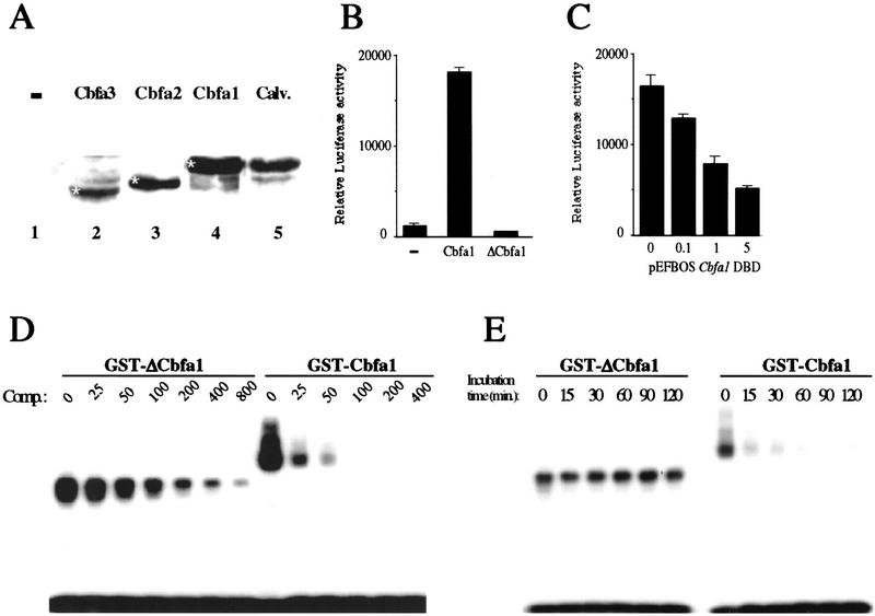
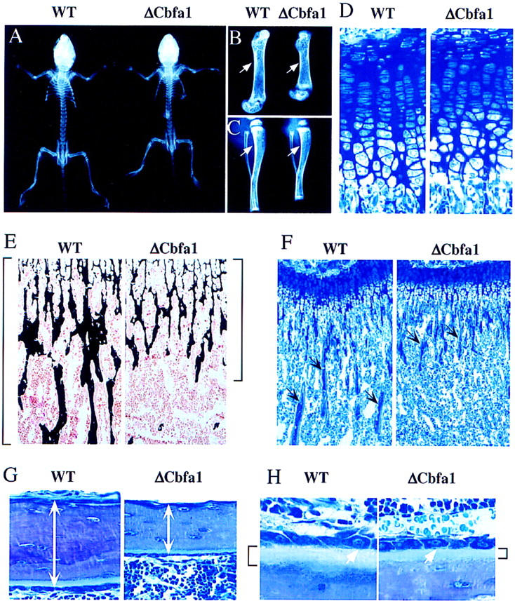
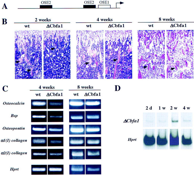
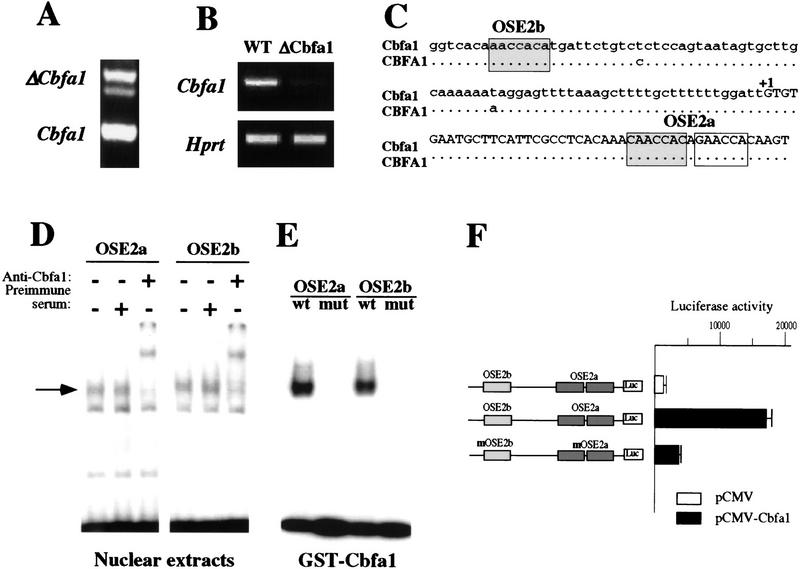
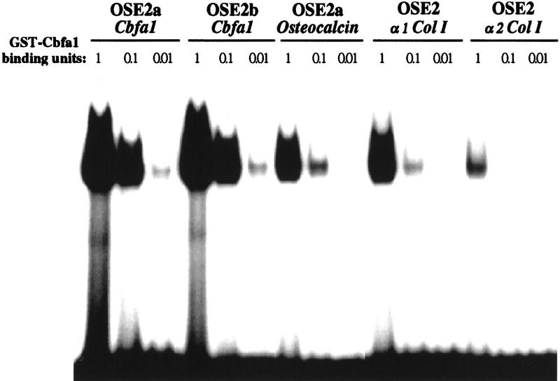
The molecular mechanisms controlling bone extracellular matrix (ECM) deposition by differentiated osteoblasts in postnatal life, called hereafter bone formation, are unknown. This contrasts with the growing knowledge about the genetic control of osteoblast differentiation during embryonic development. Cbfa1, a transcriptional activator of osteoblast differentiation during embryonic development, is also expressed in differentiated osteoblasts postnatally. The perinatal lethality occurring in Cbfa1-deficient mice has prevented so far the study of its function after birth. To determine if Cbfa1 plays a role during bone formation we generated transgenic mice overexpressing Cbfa1 DNA-binding domain (DeltaCbfa1) in differentiated osteoblasts only postnatally. DeltaCbfa1 has a higher affinity for DNA than Cbfa1 itself, has no transcriptional activity on its own, and can act in a dominant-negative manner in DNA cotransfection assays. DeltaCbfa1-expressing mice have a normal skeleton at birth but develop an osteopenic phenotype thereafter. Dynamic histomorphometric studies show that this phenotype is caused by a major decrease in the bone formation rate in the face of a normal number of osteoblasts thus indicating that once osteoblasts are differentiated Cbfa1 regulates their function. Molecular analyses reveal that the expression of the genes expressed in osteoblasts and encoding bone ECM proteins is nearly abolished in transgenic mice, and ex vivo assays demonstrated that DeltaCbfa1-expressing osteoblasts were less active than wild-type osteoblasts. We also show that Cbfa1 regulates positively the activity of its own promoter, which has the highest affinity Cbfa1-binding sites characterized. This study demonstrates that beyond its differentiation function Cbfa1 is the first transcriptional activator of bone formation identified to date and illustrates that developmentally important genes control physiological processes postnatally.


Figures

Figure 1
Figure 1
Characterization of ΔCbfa1 activity in vitro. (A) Cbfa1 is the only detectable Cbfa expressed in osteoblasts. Western blot analysis of nuclear extracts from COS cells transfected with the empty vector (−, lane 1), a Cbfa3 (lane 2), a Cbfa2 (lane 3), or a Cbfa1 (lane 3) expression vector or from calvaria (lane 5). The antiserum used recognize all three Cbfa proteins (indicated by white asteriks). (B) DNA cotransfections in COS cells. Overexpression of ΔCbfa1 does not transactivate an OSE2–luc chimeric promoter construct, whereas overexpression of Cbfa1 does. (−) cotransfection with the empty vector. (C) Osteocalcin 160-bp promoter–luc activation by endogenous Cbfa1 is inhibited by cotransfection of increasing amounts of ΔCbfa1 in ROS 17/2.8 osteoblastic cells. (DBD) DNA-binding domain. (D) Different affinity of Cbfa1 and ΔCbfa1 for OSE2. DNA competition in EMSA were performed with identical amount of GST–Cbfa1 or GST–ΔCbfa1, labeled OSE2, and increasing amounts of unlabeled OSE2 oligonucleotide (comp.). Incubation time was 5 min. (E) High stability of the ΔCbfa1–OSE2 complex. EMSA was performed with a fixed quantity of protein (GST–Cbfa1 or GST–ΔCbfa1), labeled OSE2, and a 100-fold molar excess of cold OSE2. Incubation times in presence of competitor were as indicated.
Figure 2
Figure 2
Generation of ΔCbfa1-expressing mice. (A) Representation of the ΔCbfa1 transgene. (DBD) DNA-binding domain. (B) RT–PCR showing ΔCbfa1 bone-specific expression in transgenic mice (arrow). Amplification of exon 2 of Hprt was used as an internal control for cDNA quality and PCR efficiency in each lane (bottom). (C) RT–PCR analysis comparing ΔCbfa1 level of expression in the two independent transgenic lines analyzed in this study (F21 and F28, top). RNAs were extracted from long bones of 1-month-old mice. Amplification of exon 2 of Hprt was used as an internal control for cDNA quality and PCR efficiency in each lane (bottom). PCR reactions were performed in presence of [32P]dCTP. (D) Skeletal preparation of newborn wild-type and ΔCbfa1-expressing mice. Note the presence of normally developed skull (arrowhead) and clavicles (arrow), excluding a CCD phenotype. No delayed or impaired mineralization is visible at that stage unlike the case in the Cbfa1-deficient mice (Komori et al. 1997; Otto et al. 1997).
Figure 3
Figure 3
Radiological and histological analysis of ΔCbfa1-expressing mice. (A–C) X-ray analysis of 2-week-old wild-type (WT) and ΔCbfa1-expressing mice (ΔCbfa1). ΔCbfa1-expressing mice are smaller and have shorter long bones with decreased cortical thickness (arrows). (D) Histologic analysis of the growth plate cartilage in tibia of 3-week-old wild-type and ΔCbfa1 littermates. (E–H) Histologic analysis of cancellous (E,F,H) and cortical (G) bone in 3-week-old wild-type and ΔCbfa1-expressing littermates. (E) Longitudinal sections through the tibia at the metaphysis showing mineralized trabecular bone stained in black by von Kossa and osteoid and cartilage counterstained in pink with Kernechtrot. Brackets indicate the extend of calcified bone matrix. (F) Longitudinal sections through the tibia at the metaphysis stained with toluidine blue showing the decreased amount of bone trabeculae (arrows) present in the ΔCbfa1-expressing mice. (G) Longitudinal sections through the tibia stained by toluidine blue. Cortical thickness are indicated by white arrows. (H) High magnification visualization of the osteoblasts (white arrows) present at the surface of the tibia trabeculae showing that they appear morphologically identical in 3-week-old wild-type and ΔCbfa1-expressing littermates. Note that the osteoid layer present below the osteoblasts and staining light blue (brackets) is significantly thinner in ΔCbfa1-expressing mice compared to wild-type littermates.
Figure 4
Figure 4
Bone formation and cell parameters in ΔCbfa1-expressing mice. (A) Fluorescent micrographs of the two labeled mineralization fronts at the mid-diaphysis of the tibiae of 3-week-old wild-type (WT) and ΔCbfa1-expressing mice. The brackets between the two labelings, tetracyclin at the top and calcein at the bottom, indicate the amount of newly formed bone. Note the three- to four-fold decrease in the distance between the two labeled areas in ΔCbfa1-expressing mice (B–D) Comparison of bone formation parameters in 3-week-old wild-type and ΔCbfa1-expressing mice. Measurement of bone formation rate (B), osteoid thickness (C), and trabecular bone volume (bone volume over total volume, BV/TV) (D). (E–I) Comparison of cell surfaces and numbers in 3-week-old wild-type and ΔCbfa1-expressing mice. (ObS) osteoblast surface; (BS) bone surface; (NOb) number of osteoblasts; (Bpm) bone perimeter (mm2); (OcS) osteoclast surface; (NOc) number of osteoclasts. Bars represent means ± s.e.m. (*) Statistically significant difference between wild-type and transgenic mice groups (*) P < 0.05; n = 6.
Figure 5
Figure 5
Functional analysis of the osteoblasts derived from ΔCbfa1 mice. Ex vivo culture of osteoblasts derived from calvaria of newborn wild-type and ΔCbfa1-expressing mice were analyzed after 20 days in presence of mineralization medium (see Materials and Methods). (A) Goldner trichrome staining showing that the ΔCbfa1-expressing osteoblasts form smaller and fewer nodules (arrows). (B) van Gieson staining for collagen (pink/purple color). Note the lighter aspect of the matrix secreted by the ΔCbfa1-expressing osteoblasts. (C) von Kossa staining showing that the bone matrix secreted by the ΔCbfa1-expressing osteoblasts is mineralized poorly compared to wild-type cultures. (D) Higher magnification of the culture presented in B. There are fewer and smaller collagen-secreting nodules in cultures derived from ΔCbfa1-expressing mice than from their wild-type littermates. (E) High magnification of cells stained for alkaline phosphatase. ΔCbfa1-expressing osteoblasts are synthetizing alkaline phosphatase (blue) but at a lower level than wild-type mice. (F,G) von Kossa staining for mineralized bone matrix. (F) Less mineralized nodules (arrows) are present in the ΔCbfa1-derived cultures. (G) Higher magnification of the culture presented in F, photographied in polarized light. The mineralized nodules (black) present in cultures derived from ΔCbfa1-expressing mice are smaller than the one derived from their wild-type littermates.
Figure 6
Figure 6
Osteoblast gene expression in ΔCbfa1-expressing mice. (A) RT–PCR analysis of gene expression in long bones of 2-week-old wild-type (wt) and ΔCbfa1-expressing mice. Amplification of exon 2 of Hprt was used as an internal control for cDNA quality and PCR efficiency in each lane. Expression of Osteocalcin, Bsp, and Osteopontin is virtually abolished, whereas expression of α1(I)collagen is reduced markedly, and the expresssion of α2(I)collagen is reduced moderately. (B) Northern blot analysis of α1(I)collagen expression in skin of 2-week-old wild-type and ΔCbfa1-expressing mice. The Gapdh cDNA was used to reprobe this Northern blot to assess equal loadings in the different lanes (bottom).
Figure 7
Figure 7
Auto-down-regulation of the ΔCbfa1 transgene and transient aspect of the osteopenic phenotype in ΔCbfa1-expressing mice. (A) Functional organization of the 1.3-kb OG2 promoter. The OSE1 site and the two OSE2 sites are indicated by open and solid boxes, respectively. (B) Histological analysis of long bones (tibiae) of 2-, 4-, and 8-week-old wild-type and ΔCbfa1-expressing mice. Sections through the metaphyses stained with hematoxylin/eosin. The amount of trabecular bone (pink, arrows) is severely decreased in 2-week-old trangenic mice and return to normal in 8-week-old animals. (C) RT–PCR analysis of ECM protein-encoding gene expression in long bones of 4- and 8-week-old wild-type and ΔCbfa1-expressing mice. Expression of Osteocalcin, Bone sialo protein, Osteopontin, α1(I)collagen, and α2(I)collagen in the transgenic mice is still decreased in 4-week-old animals, but less than that in 2-week-old mice (cf. with Fig. 6A), and returns to normal in 8-week-old mice. Amplification of exon 2 of Hprt was used as an internal control for cDNA quality and PCR efficiency in each lane. (D) Temporal analysis of ΔCbfa1 expression in transgenic animals. Expression of the transgene peaks at 2 weeks of age and is decreased severely thereafter. Amplification of exon 2 of Hprt was used as an internal control for cDNA quality and PCR efficiency in each lane.
Figure 8
Figure 8
Characterization of Cbfa1 autoregulation. (A) RT–PCR analysis comparing the ΔCbfa1 transgene (top) and endogenous Cbfa1 (bottom) levels of expression in long bones of 2-day-old transgenic mice. (B) Down-regulation of Cbfa1 expression in 2-week-old ΔCbfa1 compared to wild-type (WT) mice. RT–PCR analysis of gene expression in long bones of 2-week-old wild-type and ΔCbfa1-expressing mice. Amplification of exon 2 of Hprt was used as an internal control for cDNA quality and PCR efficiency in each lane. (C) Presence of three identical OSE2 elements (shaded boxes) in the promoter of the mouse and human Cbfa1 genes. (D) Binding in EMSA of ROS 17/2.8 nuclear extracts to the OSE2 elements present in Cbfa1. The specific protein–DNA complex (arrow) is supershifted by preincubation with an antiserum against Cbfa1. (E) Binding in EMSA of GST–Cbfa1 to the wild-type (wt) but not the mutated (mut) versions of the OSE2s present in Cbfa1. (F) Functional characterization of the Cbfa1 OSE2 elements. Activation of the Cbfa1 promoter containing wild-type OSE2 elements upon cotransfection with a Cbfa1 expression vector in COS cells. Mutation of the OSE2 elements inhibits this effect.
Figure 9
Figure 9
Different affinities of various OSE2s for GST–Cbfa1. Labeled OSE2s present in the Cbfa1, Osteocalcin, and type I collagen promoters were used as probes in EMSA with decreasing amount of recombinant protein as expressed by arbitrary units. A binding unit is defined as the quantity of GST–Cbfa1 required to shift 50% of the Osteocalcin promoter OSE2a probe, the initial OSE2 element described (Ducy and Karsenty 1995). GST–Cbfa1 has a 10- and 100-fold higher affinity for Cbfa1 OSE2s than for the Osteocalcin and type I collagen genes OSE2s, respectively.
Figure 10
Figure 10
Schematic representation of Cbfa1 function during and after embryonic development. (A) During embryogenesis Cbfa1 controls differentiation of a mesenchymal progenitor cell toward the osteoblastic lineage. (B) After birth, in fully differentiated osteoblasts, Cbfa1 controls the rate of bone formation by regulating directly the expression of the bone ECM-encoding genes. Moreover, Cbfa1 regulates its own gene expression.

References

    1. Amling M, Neff L, Tanaka S, Inoue D, Kuida K, Weir E, Philbrick W M, Broadus AE, Baron R. Bcl-2 lies downstream of parathyroid hormone related peptide in a signalling pathway that regulates chondrocyte maturation during skeletal development. J Cell Biol. 1997;136:205–213. - PMC - PubMed
    1. Aranson BD, Fisher AL, Blechman K, Caudy M, Gergen JP. Groucho-dependent and -independent repression activities of Runt domain proteins. Mol Cell Biol. 1997;17:5581–5587. - PMC - PubMed
    1. Aubin JE, Liu F. The osteoblast lineage. In: Bilezikian JP, Raisz LG, Rodan GA, editors. Principles of bone biology. San Diego, CA: Academic Press; 1996. pp. 51–67.
    1. Ausubel FM, Brent R, Kingston RE, Moore DD, Seidman JG, Smith JA, Struhl K. Current protocols in molecular biology. New York, NY: John Wiley & Sons; 1995.
    1. Bancroft JD, Stevens A. Theory and practice of histological techniques. New York, NY: Churchil Livingston; 1996.

Publication types

MeSH terms