. 1999 May;181(9):2773-81.
doi: 10.1128/JB.181.9.2773-2781.1999.

Glycosyltransferase domain of penicillin-binding protein 2a from Streptococcus pneumoniae is membrane associated

Affiliations

Glycosyltransferase domain of penicillin-binding protein 2a from Streptococcus pneumoniae is membrane associated

A M di Guilmi et al. J Bacteriol. 1999 May.

Abstract

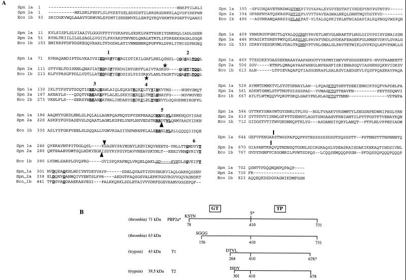
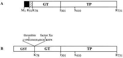
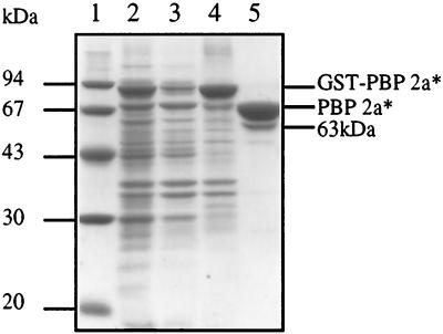
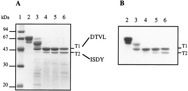
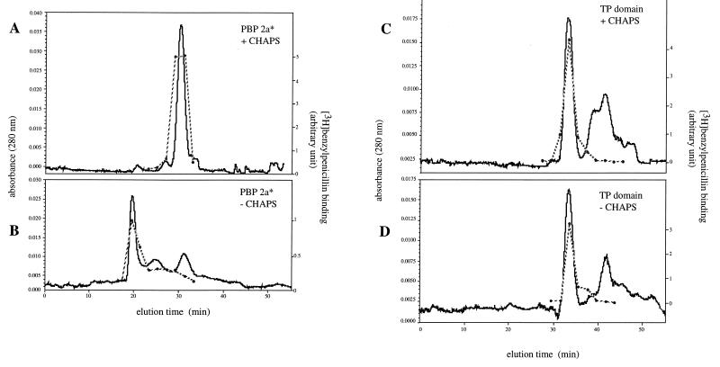
Penicillin-binding proteins (PBPs) are bacterial cytoplasmic membrane proteins that catalyze the final steps of the peptidoglycan synthesis. Resistance to beta-lactams in Streptococcus pneumoniae is caused by low-affinity PBPs. S. pneumoniae PBP 2a belongs to the class A high-molecular-mass PBPs having both glycosyltransferase (GT) and transpeptide (TP) activities. Structural and functional studies of both domains are required to unravel the mechanisms of resistance, a prerequisite for the development of novel antibiotics. The extracellular region of S. pneumoniae PBP 2a has been expressed (PBP 2a*) in Escherichia coli as a glutathione S-transferase fusion protein. The acylation kinetic parameters of PBP 2a* for beta-lactams were determined by stopped-flow fluorometry. The acylation efficiency toward benzylpenicillin was much lower than that toward cefotaxime, a result suggesting that PBP 2a participates in resistance to cefotaxime and other beta-lactams, but not in resistance to benzylpenicillin. The TP domain was purified following limited proteolysis. PBP 2a* required detergents for solubility and interacted with lipid vesicles, while the TP domain was water soluble. We propose that PBP 2a* interacts with the cytoplasmic membrane in a region distinct from its transmembrane anchor region, which is located between Lys 78 and Ser 156 of the GT domain.


Figures

FIG. 1
FIG. 1
(A) Alignment sequence of class A high-Mr PBPs S. pneumoniae PBP 1a, PBP 2a (11, 12), and E. coli PBP 1b. The conserved motifs in the GT domain are underlined and in boldface, and are numbered according to the system used in reference . The active-site conserved motifs in the TP domain are underlined. Arrowheads indicate the N-terminal positions of the PBP 1a TP domain (Ser 264) (3) and T1 and T2 tryptic products of PBP 2a (this work). The underlined residues in italics correspond to the permissive site found in E. coli PBP 1b delineating the GT and TP junction domains (16). Vertical bars indicate the C terminus of TP domains (reference and this work). Stars indicate the first residue of the detergent-free soluble proteins (reference and this work). (B) Schematic representation of the proteolytic fragments. The molecular mass of each fragment was measured by SDS-PAGE. The N-terminal sequence of the fragments was experimentally determined. The C terminus of T2 is derived from mass spectrometry measurements. The position of the active-site serine 410 is indicated (S*).
FIG. 2
FIG. 2
Schematic diagrams of S. pneumoniae PBP 2a. (A) Topology of the native PBP 2a protein. The solid and hatched boxes indicate the N-terminal cytoplasmic region and the membrane anchor, respectively. (B) Construction of the GST-PBP 2a* fusion protein. The active-site serine 410 is indicated in both figures. The peptide at the GST-PBP 2a* junction includes sequences specific for thrombin and factor Xa cleavage, as well as the polyhistidine tag. The N-terminal sequence of PBP 2a* is presented in boldface.
FIG. 3
FIG. 3
Analysis of solubilization and purification of PBP 2a*. Proteins were separated by SDS–12.5% PAGE and stained with Coomassie blue. The numbers to the left indicate the sizes of standard molecular mass markers. Lanes: 1, molecular mass markers; 2, total cell proteins; 3, soluble supernatant; 4, 0.5% CHAPS soluble extract; 5, PBP 2a* eluted from glutathione Sepharose after thrombin cleavage.
FIG. 4
FIG. 4
Trypsin cleavage of PBP 2a* leading to TP domain isolation. (A) Proteins were separated by SDS–12.5% PAGE and stained with Coomassie blue. The N-terminal sequence of each product is indicated on the right. (B) Fluorogram of a dried gel from SDS–12.5% PAGE of proteins (2 μg) labelled with [3H]benzylpenicillin prior to the migration. Digestions were performed at 37°C for 30 min. Lanes: 1, molecular mass markers; 2, undigested PBP 2a*; 3, trypsin/PBP 2a* ratio of 1:500 (wt/wt); 4, trypsin/PBP 2a* ratio of 1:100 (wt/wt); 5, trypsin/PBP 2a* ratio of 1:50 (wt/wt); 6, trypsin/PBP 2a* ratio of 1:20 (wt/wt).
FIG. 5
FIG. 5
Gel filtration of PBP 2a* and the TP domain in the presence or absence of 0.5% CHAPS. Chromatography conditions were described in Materials and Methods. Each peak aliquot was assayed for [3H]benzylpenicillin binding (dotted line). (A and B) PBP 2a*. (C and D) TP domain.
FIG. 6
FIG. 6
Charge-shift migration on native polyacrylamide gel. Proteins (5 μg) in the presence of 0.5% Triton X-100 or in a mixture of 0.5% Triton X-100–0.25% DOC were separated by native 8% PAGE and stained with Coomassie blue. Lanes: 1 to 3, PBP 2a* in 0.5% CHAPS, 0.5% Triton X-100, and 0.5% Triton X-100–0.25% DOC, respectively; 4, TP domain (star) in 0.5% Triton X-100; 5, TP domain in 0.5% Triton X-100–0.25% DOC; 6, BSA in 0.5% Triton X-100; 7, BSA in 0.5% Triton X-100–0.25% DOC.
FIG. 7
FIG. 7
Reconstitution of proteins in lipid vesicles. Proteins were reconstituted into lipid vesicles as described in Materials and Methods. The vesicles were analyzed by native 8% PAGE. (A) PBP 2a* and the TP domain. Proteins (10 μg) were stained with Coomassie blue. Lanes: 1, PBP 2a* in 3% CHAPS; 2 to 5, PBP 2a* in the presence of molar protein/lipid ratios of 0, 1/50, 1/200, and 1/500, respectively; 6, TP in 3% CHAPS; 7 to 10, TP in the presence of molar protein/lipid ratios of 0, 1/50, 1/200, and 1/500, respectively. (B) PBP 2a* thrombin-digested product. Protein detection (0.7 μg) was performed by [3H]benzylpenicillin labelling. Lanes: 1, thrombin-digested PBP 2a* in 3% CHAPS; 2 to 5, thrombin-digested PBP 2a* in the presence of molar protein/lipid ratios of 0, 1/200, 1/500, and 1/1,000, respectively.

References

    1. Adachi H, Ohta T, Matsuzawa H. A water-soluble form of penicillin-binding protein 2 of Escherichia coli constructed by site-directed mutagenesis. FEBS Lett. 1987;226:150–154. - PubMed
    1. Coffey T J, Daniels M, McDougal L K, Dowson C G, Tenover F C, Spratt B G. Genetic analysis of clinical isolates of Streptococcus pneumoniae with high-level resistance to expanded-spectrum cephalosporins. Antimicrob Agents Chemother. 1995;39:1306–1313. - PMC - PubMed
    1. Di Guilmi A M, Mouz N, Andrieu J-P, Hoskins J, Jaskunas S R, Gagnon J, Dideberg O, Vernet T. Identification, purification, and characterization of transpeptidase and glycosyltransferase domains of Streptococcus pneumoniae penicillin-binding protein 1a. J Bacteriol. 1998;180:5652–5659. - PMC - PubMed
    1. Dowson C G, Hutchison A, Spratt B G. Extensive re-modelling of the transpeptidase domain of penicillin-binding protein 2B of a penicillin-resistant South African isolate of Streptococcus pneumoniae. Mol Microbiol. 1989;3:95–102. - PubMed
    1. Ferreira L C, Schwarz U, Keck W, Charlier P, Dideberg O, Ghuysen J M. Properties and crystallization of a genetically engineered, water-soluble derivative of penicillin-binding protein 5 of Escherichia coli K12. Eur J Biochem. 1988;171:11–16. - PubMed

Publication types

MeSH terms