

Characterization of the major control region of Vibrio cholerae bacteriophage K139: immunity, exclusion, and integration
- PMID: 10217785
- PMCID: PMC93736
- DOI: 10.1128/JB.181.9.2902-2913.1999
Characterization of the major control region of Vibrio cholerae bacteriophage K139: immunity, exclusion, and integration
Abstract
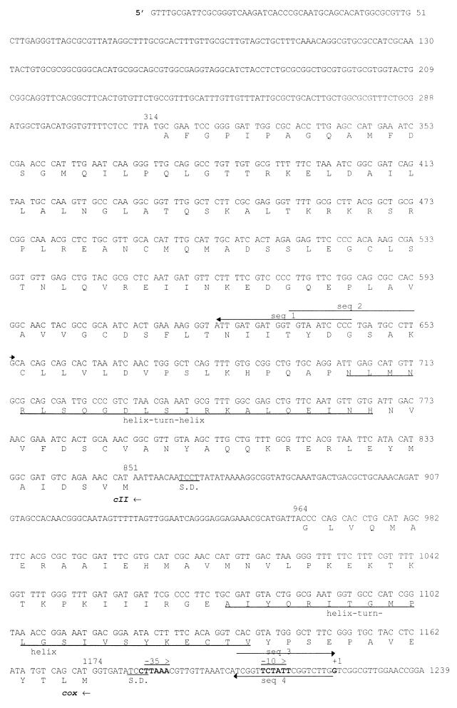
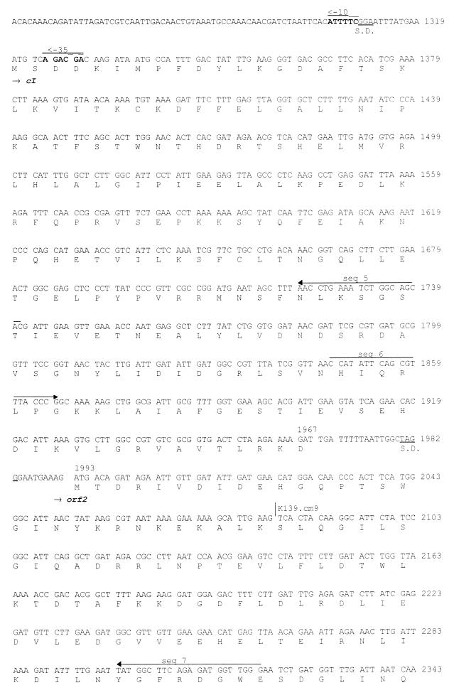
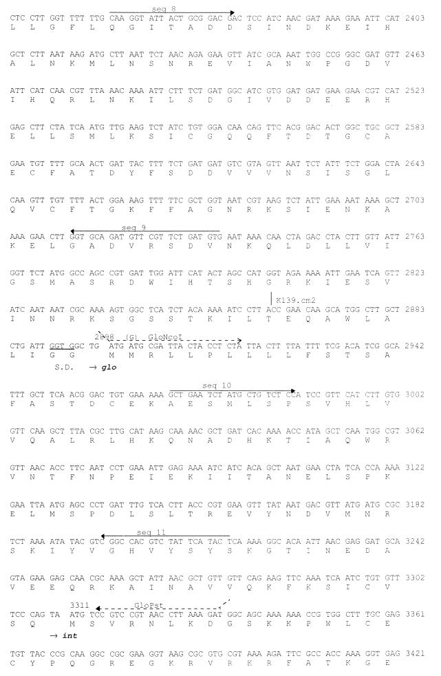
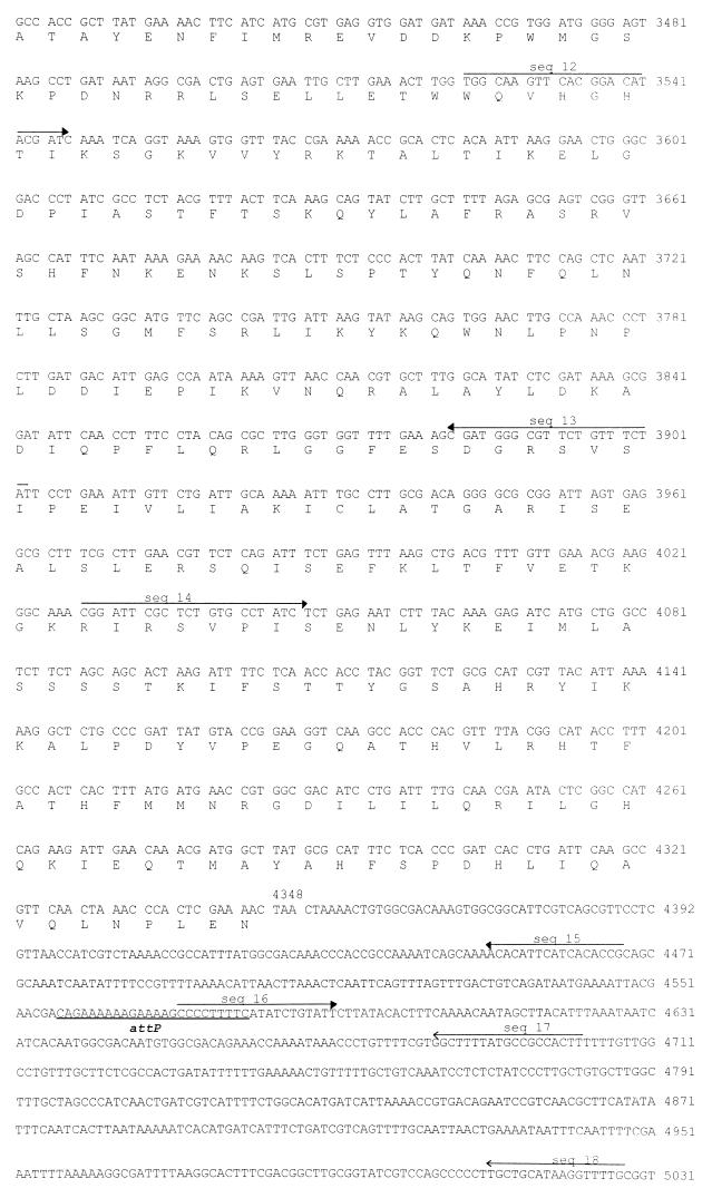
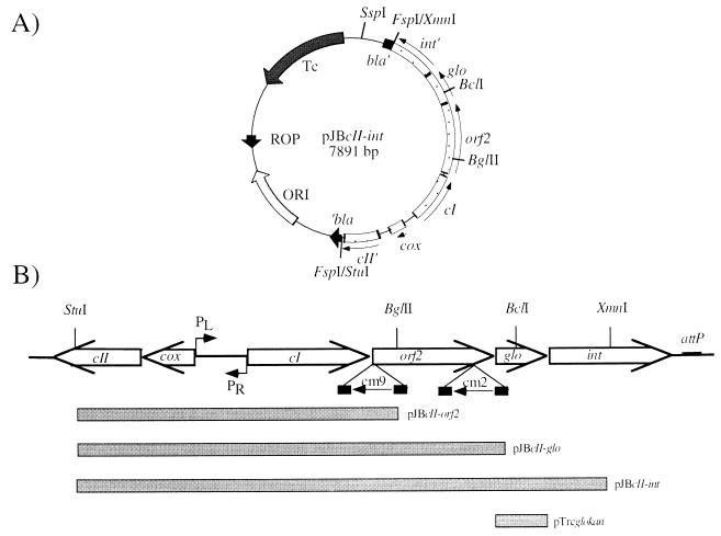
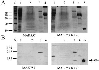
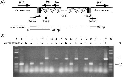
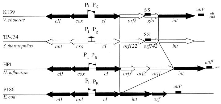
The temperate bacteriophage K139 is highly associated with pathogenic O1 Vibrio cholerae strains. The nucleotide sequence of the major control region of K139 was determined. The sequences of four (cox, cII, cI, and int) of the six deduced open reading frames and their gene order indicated that K139 is related to the P2 bacteriophage family. Two genes of the lysogenic transcript from the mapped promoter PL encode homologs to the proteins CI and Int, with deduced functions in prophage formation and maintenance. Between the cI and int genes, two additional genes were identified: orf2, which has no significant similarity to any other gene, and the formerly characterized gene glo. Further analysis revealed that Orf2 is involved in preventing superinfection. In a previous report, we described that mutations in glo cause an attenuation effect in the cholera mouse model (J. Reidl and J. J. Mekalanos, Mol. Microbiol. 18:685-701, 1995). In this report, we present strong evidence that Glo participates in phage exclusion. Glo was characterized to encode a 13.6-kDa periplasmic protein which inhibits phage infection at an early step, hence preventing reinfection of vibriophage K139 into K139 lysogenic cells. Immediately downstream of gene int, the attP site was identified. Upon analysis of the corresponding attB site within the V. cholerae chromosome, it became evident that phage K139 is integrated between the flagellin genes flaA and flaC of O1 El Tor and O139 V. cholerae lysogenic strains.
Figures
Similar articles
-
Characterization of Vibrio cholerae bacteriophage K139 and use of a novel mini-transposon to identify a phage-encoded virulence factor.Mol Microbiol. 1995 Nov;18(4):685-701. doi: 10.1111/j.1365-2958.1995.mmi_18040685.x. Mol Microbiol. 1995. PMID: 8817491
-
Vibrio cholerae phage K139: complete genome sequence and comparative genomics of related phages.J Bacteriol. 2002 Dec;184(23):6592-601. doi: 10.1128/JB.184.23.6592-6601.2002. J Bacteriol. 2002. PMID: 12426348 Free PMC article.
-
CTX prophages in classical biotype Vibrio cholerae: functional phage genes but dysfunctional phage genomes.J Bacteriol. 2000 Dec;182(24):6992-8. doi: 10.1128/JB.182.24.6992-6998.2000. J Bacteriol. 2000. PMID: 11092860 Free PMC article.
-
CTX phage of Vibrio cholerae: Genomics and applications.Vaccine. 2020 Feb 29;38 Suppl 1:A7-A12. doi: 10.1016/j.vaccine.2019.06.034. Epub 2019 Jul 1. Vaccine. 2020. PMID: 31272871 Review.
-
Survival and proliferation of the lysogenic bacteriophage CTXΦ in Vibrio cholerae.Virol Sin. 2015 Feb;30(1):19-25. doi: 10.1007/s12250-014-3550-7. Epub 2015 Jan 20. Virol Sin. 2015. PMID: 25613689 Free PMC article. Review.
Cited by
-
The vir gene of bacteriophage MAV1 confers resistance to phage infection on Mycoplasma arthritidis.J Bacteriol. 2004 Sep;186(17):5715-20. doi: 10.1128/JB.186.17.5715-5720.2004. J Bacteriol. 2004. PMID: 15317776 Free PMC article.
-
Seasonal epidemics of cholera inversely correlate with the prevalence of environmental cholera phages.Proc Natl Acad Sci U S A. 2005 Feb 1;102(5):1702-7. doi: 10.1073/pnas.0408992102. Epub 2005 Jan 14. Proc Natl Acad Sci U S A. 2005. PMID: 15653771 Free PMC article.
-
Control of directionality in integrase-mediated recombination: examination of recombination directionality factors (RDFs) including Xis and Cox proteins.Nucleic Acids Res. 2001 Jun 1;29(11):2205-16. doi: 10.1093/nar/29.11.2205. Nucleic Acids Res. 2001. PMID: 11376138 Free PMC article.
-
The Age of Phage: Friend or Foe in the New Dawn of Therapeutic and Biocontrol Applications?Pharmaceuticals (Basel). 2021 Feb 28;14(3):199. doi: 10.3390/ph14030199. Pharmaceuticals (Basel). 2021. PMID: 33670836 Free PMC article. Review.
-
Characterization of Vibrio cholerae O139 of an Aquatic Isolate in Northern Vietnam.Open Microbiol J. 2012;6:14-21. doi: 10.2174/1874285801206010014. Epub 2012 Feb 10. Open Microbiol J. 2012. PMID: 22371817 Free PMC article.
References
-
- Ackermann H W, Kasatiya S S, Kawata T, Koga T, Lee J V, Mbiguino A, Newman F S, Vieu J F, Zachary A. Classification of Vibrio bacteriophages. Intervirology. 1984;22:61–71. - PubMed
-
- Albert J B, Siddinque A K, Islam M S, Faruquwe A S G, Anzarzzaman M, Faruque S M, Sack R. Large outbreak of clinical cholera due to Vibrio cholerae non-O1 in Bangladesh. Lancet. 1993;341:704. - PubMed
-
- Altermann E, Klein J R, Henrich B. Synthesis and automated detection of fluorescently labeled primer extension products. BioTechniques. 1998;26:96–98. - PubMed
-
- Amman E, Ochs B, Abel K. Tightly regulated tac promoter vectors useful for the expression of unfused and fused proteins in Escherichia coli. Gene. 1988;69:301–315. - PubMed
Publication types
MeSH terms
Substances
Associated data
- Actions
LinkOut - more resources
Full Text Sources
