. 1999 Mar;20(3):445-56.

Segmental spinal dysgenesis: neuroradiologic findings with clinical and embryologic correlation

Affiliations

Segmental spinal dysgenesis: neuroradiologic findings with clinical and embryologic correlation

P Tortori-Donati et al. AJNR Am J Neuroradiol. 1999 Mar.

Abstract

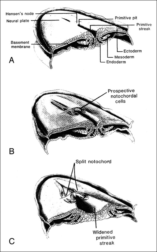
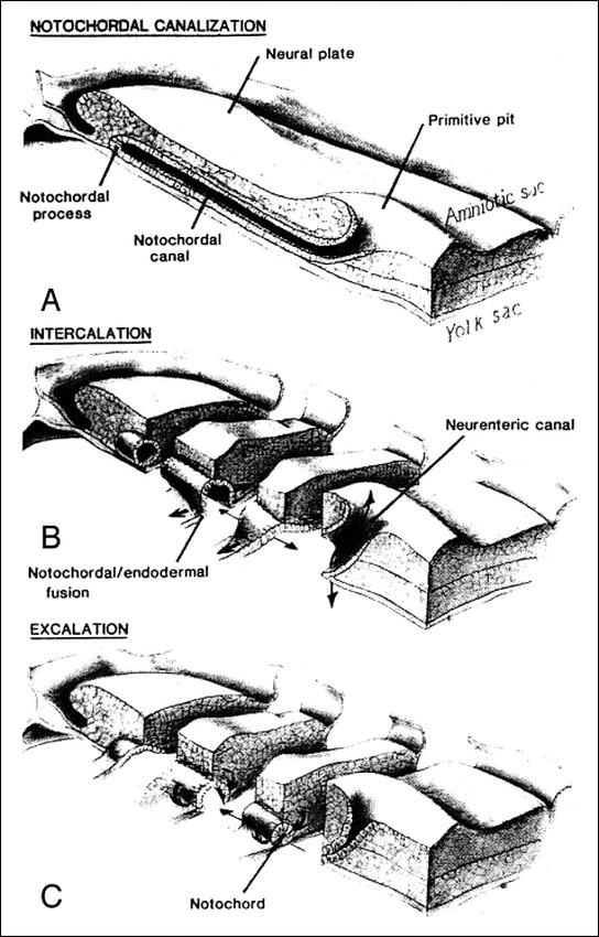
Background and purpose: Segmental spinal dysgenesis (SSD) is a rare congenital abnormality in which a segment of the spine and spinal cord fails to develop properly. Our goal was to investigate the neuroradiologic features of this condition in order to correlate our findings with the degree of residual spinal cord function, and to provide insight into the embryologic origin of this disorder. We also aimed to clarify the relationship between SSD and other entities, such as multiple vertebral segmentation defects, congenital vertebral displacement, and caudal regression syndrome (CRS).

Methods: The records of patients treated at our institutions for congenital spinal anomalies were reviewed, and 10 cases were found to satisfy the inclusion criteria for SSD. Plain radiographs were available for review in all cases. MR imaging was performed in eight patients, one of whom also underwent conventional myelography. Two other patients underwent only conventional myelography.

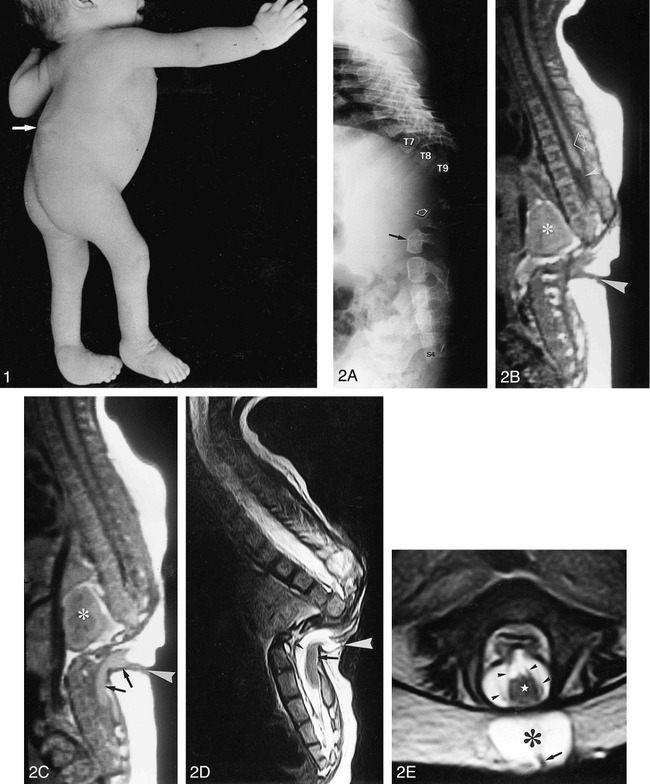
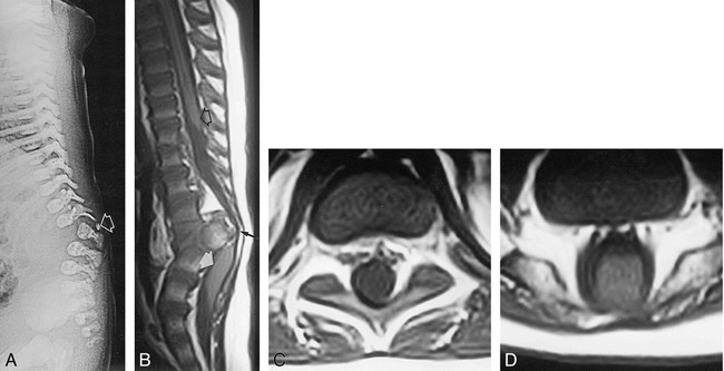
Results: Segmental vertebral anomalies involved the thoracolumbar, lumbar, or lumbosacral spine. The spinal cord at the level of the abnormality was thinned or even indiscernible, and a bulky, low-lying cord segment was present caudad to the focal abnormality in most cases. Closed spinal dysraphisms were associated in five cases, and partial sacrococcygeal agenesis in three. Renal anomalies were detected in four cases, and dextrocardia in one; all patients had a neurogenic bladder.

Conclusion: SSD is an autonomous entity with characteristic clinical and neuroradiologic features; however, SSD and CRS probably represent two faces of a single spectrum of segmental malformations of the spine and spinal cord. The neuroradiologic picture depends on the severity of the malformation and on its segmental level along the longitudinal embryonic axis. The severity of the morphologic derangement correlates with residual spinal cord function and with severity of the clinical deficit.


Figures

<sc>fig</sc> 1.
fig 1.
Case 8: 5-month-old paraplegic boy with SSD. The lower limbs are deformed, there are equinocavovarus feet, and kyphoscoliosis is present with a bony spur along the back (arrow), corresponding to the site of SSD. fig 2. Case 9: 1-month-old girl with SSD and associated partial sacrococcygeal agenesis. A, Lateral radiograph at age 12 months shows marked offset between the two spinal segments above and below the dysgenesis, resulting in marked kyphosis. Only nine thoracic vertebrae and the corresponding pair of ribs are visible; there is complete disconnection of the spine at the level of the dysgenesis, and the lower segment shows a hypoplastic L3 (open arrow) and deformed L4 (solid arrow). Partial sacrococcygeal agenesis is also present, with only S1 through S4 visible. B–E, Sagittal spin-echo (SE) T1-weighted MR images (620/30/4 [TR/TE/excitations]) at age 39 days (B and C) and sagittal fast SE T2-weighted (4000/112/2) (D) and axial fast SE T2-weighted (5000/96/2) (E) MR images at age 24 months confirm the complete disconnection of the spinal segments above and below the dysgenesis. A horseshoe kidney is seen in B and C (asterisk). The upper cord ends at the midthoracic level with a wedge-shaped termination (open arrow, B); a questionable threadlike structure extends below (solid arrow, B). No cord is discernible at the level of the anomaly. Below, the bulky, low-lying lower cord (arrows, C and D) appears to connect with the bottom of a dermal sinus (white arrowhead, B–D). Nerve roots with an aberrant course anteriorly to the lower cord are seen on T2-weighted images (black arrowhead, D). Note worsening of the degree of kyphosis at age 24 months (compare C and D), causing the dermal sinus to collapse. In the axial plane, the bulky lower cord (star, E) and nerve roots are visible coursing anterior to the cord (black arrowheads, E). Posteriorly, the CSF-filled lower spinal canal (asterisk, E) connects with the dermal sinus (arrow, E).
<sc>fig</sc> 3.
fig 3.
Case 10: 2-month-old girl with SSD and associated questionable coccygeal agenesis. A, Lateral radiograph at age 1 day shows severe hypoplasia and posterior dislocation of L2 (arrow) with absence of posterior vertebral elements. There is marked kyphosis in the upper lumbar spine and questionable absence of the coccyx, as its ossification may be incomplete in the neonate. B–D, Sagittal SE (600/20/4) (B) and axial SE (600/16/2) (C and D) T1-weighted MR images at age 12 months show that the upper cord ends at the midthoracic level in a wedge shape (open arrow, B) and contains a thin hydromyelic cavity. Below, no cord is discernible even on the axial images (C and D). At the apex of the kyphosis, the spinal canal is extremely narrowed (black arrow, B). Below, a bulky, low-lying lower cord is visible in the thecal sac (white arrow, B) and appears to fill it almost completely (D).
<sc>fig</sc> 4.
fig 4.
Case 1: 8-month-old boy. A and B, Sagittal SE T1-weighted MR image (500/14/2) (A) and 3-mm-thick reformatted sagittal MR image obtained from a 3D gradient-echo sequence (20/7/1) with 30° flip angle (80 original 1-mm-thick partitions) (B) at age 9 years. In this case the malformation involves the upper lumbar segment. The degree of kyphosis is moderate, and there is no disconnection of the spinal canal. The upper cord (open arrow, A) is continuous with a hypoplastic cord segment (arrowheads, A). Below, the lower cord (solid arrow, A) is bulky and low-lying. The abrupt change in caliber of the spinal cord may be seen more easily on the reformatted image (arrowheads, B). fig 5.  Case 5: 5-month-old boy with SSD involving the lumbosacral spine. Sagittal SE T1-weighted (600/20/4) MR image at age 2 years shows marked kyphosis in the lumbosacral spine with absent or undetermined vertebrae. The spinal cord is hypoplastic and blends with the filum terminale, surrounding the dysgenetic vertebral bodies and apparently tethering to the sacrum (arrowheads). Because the dysgenesis involves the lowermost portion of the neural plate, there cannot be a lower cord segment; in fact, this particular case illustrates an overlap between SSD and CRS. There is concurrent hydromyelia in the thoracic spinal cord (arrows).
<sc>fig</sc> 6.
fig 6.
A, Normal human gastrulation. Prospective endodermal and mesodermal cells of the epiblast migrate toward the midline and ingress (arrows) through defects in the basement membrane at the primitive streak to become definitive endoderm and mesoderm. B, Normal human gastrulation. Prospective notochordal cells located along the anterior margin of Hensen's node ingress through the primitive pit to become the notochordal process located between the ectoderm and endoderm. C, Abnormal gastrulation resulting in a failure of midline axial integration. The primitive streak is abnormally wide; prospective notochordal cells therefore begin ingression more laterally than normal. As a result, two notochordal processes are formed. The caudal neuroepithelium, comprising two columns of tissue that flank and are separated by the primitive streak, fails to become integrated to form a single neuroepithelial sheet and instead forms two hemineural plates. The type of resultant malformation depends on the level and extent of the abnormality, and the success of subsequent reparative efforts. Reproduced from (22) with permission of S. Karger AG, Basel, Switzerland.
<sc>fig</sc> 7.
fig 7.
Notochordal canalization, intercalation and excalation. A, The notochordal process contains a central lumen (the notochordal canal), which is continuous with the amniotic cavity through the primitive pit. B, During intercalation, the canalized notochordal process fuses with the underlying endoderm; the communication of the amnion with the yolk sac forms the primitive neurenteric canal. C, During excalation, the notochord rolls up and separates from the endoderm to become the definitive notochord; the primitive neurenteric canal becomes obliterated. Reproduced from with permission of S. Karger AG, Basel, Switzerland.

Similar articles

Cited by

References

    1. Scott RM, Wolpert SM, Bartoshesky LF, Zimbler S, Karlin L. Segmental spinal dysgenesis. Neurosurgery 1988;22:739-744 - PubMed
    1. Faciszewski T, Winter RB, Lonstein JE, Sane S, Erickson D. Segmental spinal dysgenesis: a disorder different from spinal agenesis. J Bone Joint Surg [Am] 1995;77:530-537 - PubMed
    1. Fondelli MP, Cama A, Rossi A, Piatelli GL, Tortori-Donati P. Segmental spinal dysgenesis: MRI findings in 7 cases (abstr). Neuroradiology 1996;38(Suppl 2):89
    1. Raybaud CA, Couallier JF, Choux M, Binnert D. The “interrupted cord”: three cases of an unusual malformation of the spinal cord (abstr). AJNR Am J Neuroradiol 1988;9:1007
    1. McKay DW, Nason SS. Congenital duplication of the spinal canal. Spine 1980;5:390-391 - PubMed

MeSH terms