. 1999 Apr 27;96(9):4814-9.
doi: 10.1073/pnas.96.9.4814.

Identification of a photoreceptor cell-specific nuclear receptor

Affiliations

Identification of a photoreceptor cell-specific nuclear receptor

M Kobayashi et al. Proc Natl Acad Sci U S A. .

Abstract

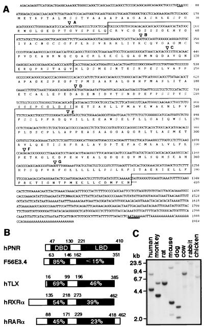
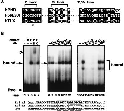
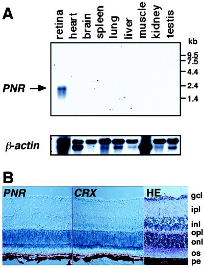
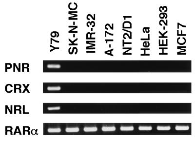
Nuclear receptors comprise a large and expanding family of transcription factors involved in diverse aspects of animal physiology and development, the functions of which can be modulated in a spatial and temporal manner by access to small lipophilic ligands and/or the specificity of their own localized expression. Here we report the identification of a human nuclear receptor that reveals a unique proximal box (CNGCSG) in the DNA-binding domain. The conservation of this feature in its nematode counterpart suggests the requirement for this type of P box in the genetic cascades mediated by nuclear receptors in a wide variety of animal species. The expression of this receptor, PNR (photoreceptor-specific nuclear receptor), appears strongly restricted in the retina, exclusively in photoreceptor cells. In human cell lines, PNR expression was observed in Y79 retinoblastoma along with other photoreceptor marker genes such as CRX. Among vertebrate receptors, PNR shares structural kinship with an orphan receptor TLX, and despite distinct differences in the DNA binding domain, PNR is able to recognize a subset of TLX target sequences in vitro. Analyses of the human PNR gene revealed its chromosomal position as 15q24, a site that has recently been reported as a susceptible region for retinal degeneration. These data support a role for PNR in the regulation of signalling pathways intrinsic to the photoreceptor cell function.


Figures

Figure 1
Figure 1
(A) The human PNR cDNA and predicted amino acid sequences. An in-frame stop codon preceding the initiation methionine codon is underlined. DBD and LBD regions are boxed. There are some sequence ambiguities in the 3′ untranslated region. A potential poly(A)+ signal (AATAAA) is double-underlined. Intron positions are indicated by ▿ (see Table 1 for details). (B) Comparison of human PNR (hPNR) with nematode F56E3.4 and other nuclear receptors. The % identity is indicated in the aligned sequences for the DBDs and LBDs. (C) Southern blot analysis using the LBD region of the human PNR cDNA as a probe. Distinct bands were dectected in all lanes.
Figure 2
Figure 2
(A) Alignment of the amino acid residues in the P box, D box and T/A box regions of the DBDs between human PNR, C. elegans F56E3.4 and human TLX. Conserved residues are shaded. Gaps are indicated by a dashed line. ∗ indicates the position of the serine residue in the P boxes of these 3 receptors that replaces an otherwise strictly conserved lysine. (B) In vitro DNA binding specificity by PNR protein. Lanes 1–5 show DNA-binding patterns of COS cell-expressed HA-tagged PNR protein to Kni x2 probe. Lane 1 is probe only, and mock cell extract was added in lane 2. Lane 3 shows a specific protein–DNA complex formed when cell extract containing HA-hPNR is added. This band is supershifted by addition of antibody against the HA tag (lane 4) but is not affected by nonspecific antibody (lane 5). Lanes 6–12 show that PNR bound to labeled Kni x2 probe can be blocked by competition by addition of excess Kni x2 cold probes (lane 8), but not by Kni x1, which is based on the TLL binding site identified in the D. melanogaster knirps gene (17), or 3MT probes (lanes 9–12). Competitor probes are indicated above, with 10-fold excess added in lanes 7, 9, and 11 and 50-fold excess in lanes 8, 10, and 12. Lanes 13–20 show that COS cell-expressed chicken TLX protein forms two distinct complexes on the Kni x2 probe (lane 14), both of which can be efficiently blocked by competition by either Kni x2 or Kni x1, but not the mutated 3MT probe.
Figure 3
Figure 3
Expression of PNR mRNA in rodents. (A) Northern hybridization analyses of mRNA from rat tissues using the human PNR cDNA as a probe. All lanes contain 2 μg of poly(A)+ RNA. β-Actin was used to normalize for RNA quality. (B) In situ hybridization of mouse retina sections with digoxigenin-labeled PNR and CRX RNA probes. An adjacent section was stained with hematoxylin/eosin (HE) to help visualize different cell layers. gcl, ganglion cell layer; ipl, inner plexiform layer; inl, inner nuclear layer; opl, outer plexiform layer; onl, outer nuclear layer; os, outer segments; pe, pigment epithelium.
Figure 4
Figure 4
Expression of PNR, CRX, NRL, and RARα in human cell lines. RT-PCR analysis of total RNA isolated from various human cell lines. Y79, retinoblastoma; SK-N-MC and IMR-32, neuroblastoma; A-172, glioblastoma; NT2/D1, embryonic carcinoma; HeLa, cervical carcinoma; HEK-293, adenovirus-transformed embryonic kidney fibroblast; and MCF7, mammary tumor.
Figure 5
Figure 5
Primary structure of the 5′ upstream region of the human PNR gene. The end of the longest 5′ RACE clones is indicated by ∗ (denoted as +1 in the numbering of the nucleotides) and the upstream TATA box (CATAAA) is shown (boxed). The 5′ end of the cDNA in Fig. 1A is indicated (†). The first ATG is in bold, with an in-frame upstream stop codon underlined. The junction between the first and second exons is shown by the arrow, and the positions of the primers used in the 5′ RACE analysis are marked and labeled. A CRX consensus binding sequence just upstream of the TATA box is indicated by double-underline.
Figure 6
Figure 6
Chromosomal localization of the human PNR gene. (Left) A representative fluorescence in situ hybridization pattern obtained with the human PNR P1 genomic clone as a probe. (Right) The same metaphase chromosome preparation stained with 4′,6-diaminido-2-phenylindole (DAPI). The human PNR gene was assigned to 15q24 (arrowheads).

References

    1. Enmark E, Gustafsson J A. Mol Endocrinol. 1996;10:1293–1307. - PubMed
    1. Blumberg B, Evans R M. Genes Dev. 1998;12:3149–3155. - PubMed
    1. Willy P J, Mangelsdorf D J. Horm Signal. 1998;1:307–358.
    1. Escriva H, Safi R, Hanni C, Langlois M C, Saumitou-Laprade P, Stehelin D, Capron A, Pierce R, Laudet V. Proc Natl Acad Sci USA. 1997;94:6803–6808. - PMC - PubMed
    1. Yu R T, McKeown M, Evans R M, Umesono K. Nature (London) 1994;370:375–379. - PubMed

Publication types

MeSH terms

Substances

Associated data