. 1999 Apr 27;96(9):4971-6.
doi: 10.1073/pnas.96.9.4971.

Rapid pole-to-pole oscillation of a protein required for directing division to the middle of Escherichia coli

Affiliations

Rapid pole-to-pole oscillation of a protein required for directing division to the middle of Escherichia coli

D M Raskin et al. Proc Natl Acad Sci U S A. .

Abstract

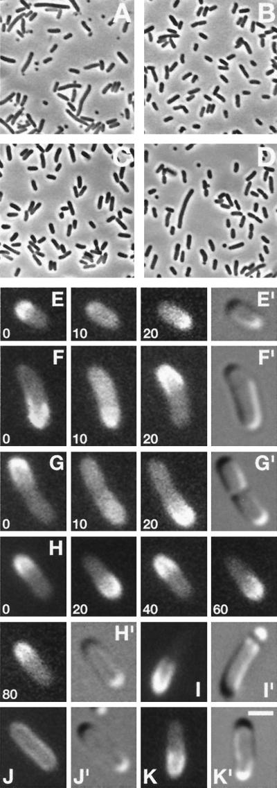
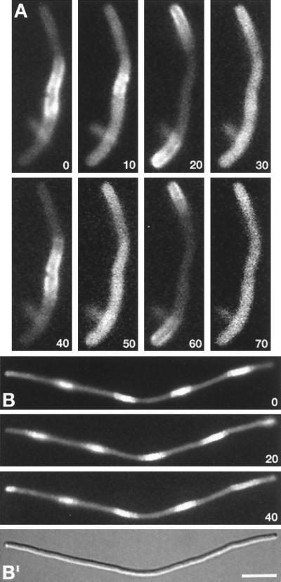
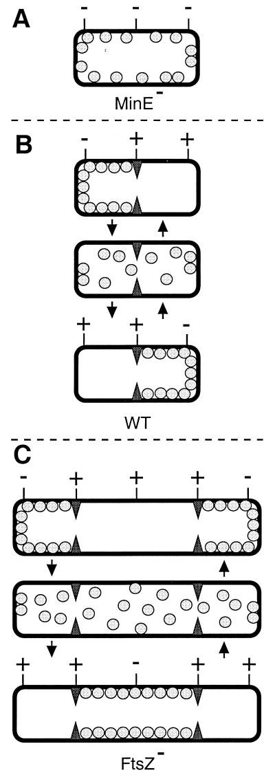
Accurate placement of the division septum at the midpoint of Escherichia coli cells requires the combined action of a general division inhibitor (MinC), a site-specific suppressor of division inhibition (MinE), and an ATPase (MinD) that is required for proper functioning of both MinC and MinE. We previously showed that a functional MinE-Gfp fusion accumulates in a ring structure at/near the middle of cells. Here we show that functional Gfp-MinD accumulates alternately in either one of the cell halves in what appears to be a rapidly oscillating membrane association-dissociation cycle imposed by MinE. The results indicate that MinD represents a novel type of dynamic cellular element in bacteria, with multiple roles in directing the division apparatus to the middle of the cell.


Figures

Figure 1
Figure 1
Dynamic properties of functional Gfp-MinD in live cells. Phase (AD), fluorescence (EK), and differential interference contrast (E′–K′) micrographs showing properties of Gfp-MinD. Cells were grown either in the absence (A and C) or presence of 25 μM (D and I), or 37 μM (B, EH, J, and K) IPTG. (A and B) Correction of minD1 by gfp-minD in strain DR104(λDR119) [minD1 recA∷Tn10 (Placgfp-minD)]. (C and D) The induction of minicell formation by moderate overexpression of Gfp-MinD in strain PB103/pDR119 (wt/Placgfp-minD). (EH) Time-lapse images showing Gfp-MinD oscillation in normally dividing cells of strains DR104(λDR122) [minD1 recA∷Tn10 (Placgfp-minDE)] (F and G), and PB103(λDR122) [wt(Placgfp-minDE)] (E and H). Times are indicated in sec. (I) Gfp-MinD segregation and minicell formation in a PB103/pDR119 cell. (J and K) Gfp-MinD localization in strain PB114 (ΔminCDE) lysogenic for either λDR119 (Placgfp-minD) (J) or λDR122 (Placgfp-minDE) (K). [Bar represents 1 μm (EK) or 4 μm (AD).]
Figure 2
Figure 2
Gfp-MinD localization in FtsZ filaments. Time-lapse fluorescence images of a short (A) and long (B) filament of strain DR102(λDR122)/pDB346 [ΔminCDEaph ftsZ0 recA∷Tn10 (Placgfp-minDE)/PλRftsZ cI857]. Times are indicated in sec. (B′) A differential interference contrast image of the filament in B. Cells were grown at 30°C (resulting in repression of ftsZ expression) in the presence of 37 μM IPTG. [Bar represents 2.5 μm (A) or 5.0 μm (B).]
Figure 3
Figure 3
Model for MinD and MinE action in preventing aberrant septation events. MinD is represented by gray spheres, the MinE ring by filled triangles, and PDSs by either a − (blocked by MinC/MinD action) or + (not blocked by MinC/MinD, available for assembly of FtsZ ring) sign. (A) In the absence of MinE, MinD is distributed evenly over the membrane. Provided MinC is present, this prevents septal ring formation at all PDSs, resulting in the formation of nonseptate filaments (7). (B) In WT cells, MinD oscillates from one side of the MinE ring to the other, alternately blocking division at each of the polar PDSs. For simplicity, it is assumed that the MinC/MinD division block is relieved as soon as MinD leaves a PDS, although it may well remain refractive to FtsZ assembly for some period afterward. (C) In the absence of FtsZ, multiple MinE rings define three or more cell segments. As in WT cells, MinD oscillates between the segments flanking each MinE ring.
Figure 4
Figure 4
Identification of Gfp-MinD with MinD-specific antiserum. Immunoblot showing Gfp-MinD (58.1 kDa, upper arrow) and native MinD (29.6 kDa, lower arrow) as detected with MinD-specific antiserum. Cells were grown in the presence of 37 μM (lanes 1–5) or 25 μM (lanes 6–9) IPTG to an OD (600 nm) of 0.3, and whole-cell extracts were prepared. Lanes 1–5 contained 20 μg total protein of strains PB114 [ΔminCDE] (lane 1); PB103(λDR119) [wt(Placgfp-minD)] (lane 2); PB103(λDR122) [wt(Placgfp-minDE)] (lane 3); PB114(λDR119) [ΔminCDE (Placgfp-minD)] (lane 4); and PB114(λDR122) [ΔminCDE (Placgfp-minDE)] (lane 5). Lanes 6–8 contained, respectively, 4, 2, and 1 μg of PB103/pDR119 (wt/Placgfp-minD), and lane 9 contained 2 μg of PB103/pDR122 (wt/Placgfp-minDE). Samples in lanes 6–9 were mixed with appropriate amounts of PB114 extract such that each lane contained 20 μg of total protein.

Comment in

  • Bacterial cell division: a moveable feast.
    Jacobs C, Shapiro L. Jacobs C, et al. Proc Natl Acad Sci U S A. 1999 May 25;96(11):5891-3. doi: 10.1073/pnas.96.11.5891. Proc Natl Acad Sci U S A. 1999. PMID: 10339512 Free PMC article. Review. No abstract available.

References

    1. Bramhill D. Annu Rev Cell Dev Biol. 1997;13:395–424. - PubMed
    1. Lutkenhaus J, Addinall S G. Annu Rev Biochem. 1997;66:93–116. - PubMed
    1. Margolin W. Trends Microbiol. 1998;6:233–238. - PubMed
    1. Rothfield L I, Justice S S. Cell. 1997;88:581–584. - PubMed
    1. Adler H I, Fisher W D, Cohen A, Hardigree A A. Proc Natl Acad Sci USA. 1967;57:321–326. - PMC - PubMed

Publication types

MeSH terms

LinkOut - more resources