. 1999 Apr 27;96(9):4977-82.
doi: 10.1073/pnas.96.9.4977.

Identification of profilin and src homology 3 domains as binding partners for Drosophila enabled

Affiliations

Identification of profilin and src homology 3 domains as binding partners for Drosophila enabled

S M Ahern-Djamali et al. Proc Natl Acad Sci U S A. .

Abstract

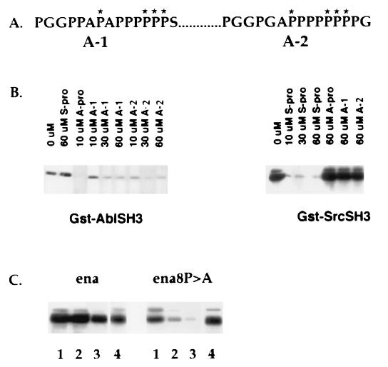
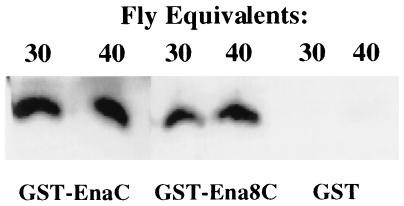
Drosophila Enabled (Ena) was first identified as a genetic suppressor of mutations in the Abelson tyrosine kinase and subsequently was shown to be a member of the Ena/vasodilator-stimulated phosphoprotein family of proteins. All members of this family have a conserved domain organization, bind the focal adhesion protein zyxin, and localize to focal adhesions and stress fibers. Members of this family are thought to be involved in the regulation of cytoskeleton dynamics. The Ena protein sequence has multiple poly-(L-proline) residues with similarity to both profilin and src homology 3 binding sites. Here, we show that Ena can bind directly to the Drosophila homolog of profilin, chickadee. Furthermore, Ena and profilin were colocalized in spreading cultured cells. We report that the proline-rich region of Ena is responsible for this interaction as well as for mediating binding to the src homology 3 domain of the Abelson tyrosine kinase. These data support the hypothesis that Ena provides a regulated link between signal transduction and cytoskeleton assembly in the developing Drosophila embryo.


Figures

Figure 1
Figure 1
Identification of chickadee as a binding partner for Ena in a yeast two-hybrid screen. (A) The first 235 amino acids of Ena (EnaN) and last 243 amino acids of Ena (EnaC) were fused to the sequence encoding the GAL4 DNA binding domain in the pAS1-CYH2 vector. These two constructs were cotransformed with GAL4AD-chickadee (Chic), a clone in which the full-length chickadee coding sequences were fused to the sequence encoding the GAL4 activation domain and which was isolated in a yeast two-hybrid screen by using the C-terminal 243 amino acids of Ena as bait. From each of the cotransformations, two independent clones were spread on medium lacking histidine and analyzed for growth. EnaC, which contains two putative profilin binding sites, can interact with chickadee, whereas EnaN, which contains no putative binding sites for profilin, cannot. (B). Amino acids 440–490 from EnaC containing the putative profilin binding sites (EnaC/Pro) and amino acids 490–684 from EnaC lacking any putative profilin binding sites (EnaC/EVH2) were fused to the DNA binding domain of the yeast transcription factor GAL4 and cotransformed with GAL4AD chickadee. Cotransformed yeast was spread on medium lacking histidine and analyzed for growth. EnaC/Pro can interact with chickadee, whereas EnaC/EVH2 cannot.
Figure 2
Figure 2
Binding of Ena protein to the Drosophila profilin homolog, chickadee. Serial dilutions of whole adult Drosophila lysates were split into two equal aliquots and bound in solution to an equal amount of either GST alone or GST fused to Ena amino acids 374–684. Retained proteins after solution binding were detected with anti-chickadee antibodies. The GST–Ena protein bound reproducibly to chickadee. The faint band seen in the GST alone bands is nonspecific Ena binding to the GST control.
Figure 3
Figure 3
Colocalization of Ena and profilin in cultured spreading PTK2 cells. PTK2 cells were transfected with an expression vector carrying the ena cDNA. On the second day after transfection, cells were treated with trypsin and allowed to spread on coverslips for 30 min. Cells were fixed and made permeable in methanol and double-labeled for Ena (A) and profilin (B). C merges A and B. Both Ena and profilin can be detected at the periphery of the spreading cells. (Bar = 20 μm.)
Figure 4
Figure 4
SH3 domain binding of Ena. S2 cells were transfected with Ena under the control of an actin promoter. Proteins retained after solution binding to equal amounts of various GST–SH3 domains were blotted with anti-Ena N-terminal antibodies. None of the proteins bound to the GST negative control (data not shown). Ena binds to the murine and Drosophila Abl SH3 domains, the src SH3 domain, and the C-terminal SH3 domain of Drk.
Figure 5
Figure 5
Proline-rich motifs in Ena involved in SH3 binding. (A) A-1 represents Ena amino acids 401–415, and A-2 represents Ena amino acids 464–478. Asterisks indicate the residues chosen for mutagenesis from proline to alanine to generate Ena8. (B) S2 cells were transfected with Ena, and the lysates were incubated with glutathione-Sepharose-bound GST–AblSH3 or SrcSH3 preincubated with the indicated concentrations of peptides. Bound proteins were blotted with anti-Ena antibodies. The optimal consensus binding motifs for Abl (Abl–Pro) and Src (Src–Pro) specifically blocked binding of Ena to the Abl or to the Src SH3 domains, respectively. Ena A-1 at 30 μM and Ena A-2 at 10 μM specifically blocked binding of Ena to the AblSH3 domain. (C) Wild-type ena or ena8 was transfected into S2 cells. Serial 2-fold dilutions of transfected cell lysates were incubated with glutathione-Sepharose-bound GST–AblSH3 (lanes 1–3). Bound proteins were blotted with anti-Ena antibodies. Binding of the mutant Ena8 to the Abl SH3 domain was significantly reduced. The difference in binding between wild-type Ena and Ena8 was more evident at lower concentrations of protein. Expression levels of wild-type and mutant Ena were equivalent (lane 4).
Figure 6
Figure 6
Chickadee binding to the Ena mutant protein Ena8 P → A. Whole Drosophila lysates from 60 or 80 adult flies were split into three equal aliquots and bound in solution to equal amounts of GST alone, GST fused to Ena amino acids 374–684, or GST fused to Ena8 P → A amino acids 374–684. Proteins retained after solution binding were detected with anti-chickadee antibodies. Reproducibly, the GST–Ena protein, the GST–Ena8 P → A protein, and GST alone bound equally well to chickadee.

References

    1. Carlier M F. Curr Opin Cell Biol. 1998;10:45–51. - PubMed
    1. Dramsi S, Cossart P. Annu Rev Cell Dev Biol. 1998;14:137–166. - PubMed
    1. Elch M D, Mallavarapu A, Rosenblat J, Mitchison T J. Curr Opin Cell Biol. 1997;9:54–61. - PubMed
    1. Ahern-Djamali S M, Comer A R, Bachmann C, Kastenmeier A S, Reddy S K, Beckerle M C, Walter U, Hoffmann F M. Mol Biol Cell. 1998;9:2157–2171. - PMC - PubMed
    1. Gertler F, Niebuhr K, Reinhard M, Wehland J, Soriano P. Cell. 1996;87:227–239. - PubMed

Publication types