. 1999 Apr 27;96(9):5007-12.
doi: 10.1073/pnas.96.9.5007.

The intracellular domain of interferon-alpha receptor 2c (IFN-alphaR2c) chain is responsible for Stat activation

Affiliations

The intracellular domain of interferon-alpha receptor 2c (IFN-alphaR2c) chain is responsible for Stat activation

S V Kotenko et al. Proc Natl Acad Sci U S A. .

Abstract

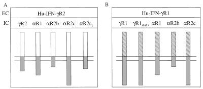
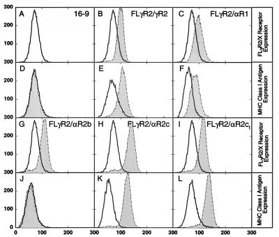
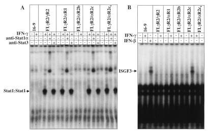
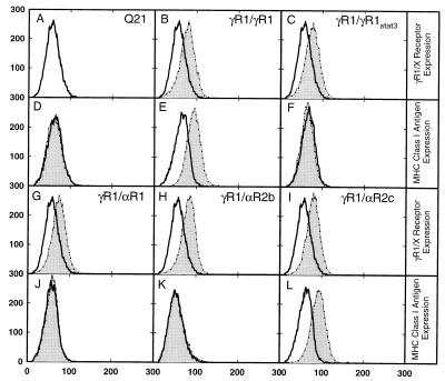
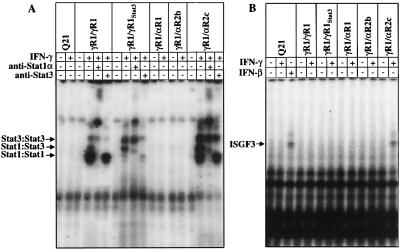
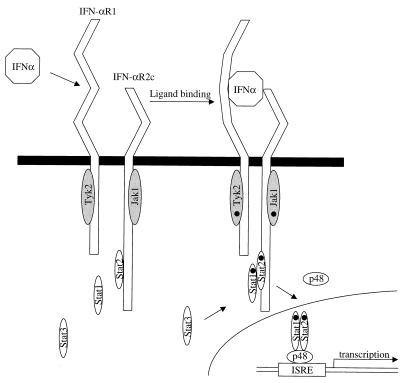
Type I IFNs activate the Jak-Stat signal transduction pathway. The IFN-alpha receptor 1 (IFN-alphaR1) subunit and two splice variants of the IFN-alphaR2 subunit, IFN-alphaR2c and IFN-alphaR2b, are involved in ligand binding. All these receptors have been implicated in cytokine signaling and, specifically, in Stat recruitment. To evaluate the specific contribution of each receptor subunit to Stat recruitment we employed chimeric receptors with the extracellular domain of either IFN-gammaR2 or IFN-gammaR1 fused to the intracellular domains of IFN-alphaR1, IFN-alphaR2b, and IFN-alphaR2c. These chimeric receptors were expressed in hamster cells. Because human IFN-gamma exhibits no activity on hamster cells, the use of the human IFN-gamma receptor extracellular domains allowed us to avoid the variable cross-species activity of the type I IFNs and eliminate the possibility of contributions of endogenous type I IFN receptors into the Stat recruitment process. We demonstrate that Stat recruitment is solely a function of the IFN-alphaR2c intracellular domain. When chimeric receptors with the human IFN-gammaR1 extracellular domain and various human IFN-alpha receptor intracellular domains were expressed in hamster cells carrying the human IFN-gammaR2 subunit, only the IFN-alphaR2c subunit was capable of supporting IFN-gamma signaling as measured by MHC class I induction, antiviral protection, and Stat activation. Neither the IFN-alphaR2b nor the IFN-alphaR1 intracellular domain was able to recruit Stats or support IFN-gamma-induced biological activities. Thus, the IFN-alphaR2c intracellular domain is necessary and sufficient to activate Stat1, Stat2, and Stat3 proteins.


Figures

Figure 1
Figure 1
Structure of chimeric receptors. The Hu-IFN-γR1/γR1 and Hu-IFN-γR2/γR2 are the first (15) and second (16) chains of the Hu-IFN-γ receptor complex, where the NheI site was introduced in the beginning of the transmembrane domain of these receptors. The extracellular domain (EC) of either Hu-IFN-γR1 (B, hatched bars) or Hu-IFN-γR2 (A, open bars) was fused to the transmembrane and intracellular domains (IC) of different subunits of the Hu-IFN-α receptor complex (shaded bars): the Hu-IFN-αR1 (9); the Hu-IFN-αR2b (10), and the Hu-IFN-aR2c (11, 12). The FLAG epitope was introduced at the NH2 terminus of the Hu-IFN-γR2 extracellular domain. The FLγR2/αR2ct chimera has the IFN-αR2c intracellular domain prematurely terminated after Asp-315. The γR1/γR1Stat3 chimera has the Stat1α recruitment site replaced by the Stat3 recruitment site.
Figure 2
Figure 2
Expression of receptors on the cell surface and induction of HLA-B7 surface antigen in hamster 16-9 cells by IFN-γ. The expression of FLγR2/γR2, FLγR2/αR1, FLγR2/αR2b, FLγR2/αR2c, and FLγR2/αR2ct (B, C, G, H, and I) or induction of HLA-B7 antigen by IFN-γ (D, E, F, J, K, and L) were analyzed by flow cytometry. Ordinate, relative number of cells; abscissa, relative fluorescence. (B, C, G, H, and I) Cells were harvested and incubated with anti-FLAG monoclonal antibody (thin lines, shaded areas), the parental 16-9 cells were used as a control (A, B, C, G, H, and I, thick lines, open areas). (D, E, F, J, K, and L) Cells were treated with IFN-γ (thin lines, shaded areas) or left untreated (thick lines, open areas). Cells were the parental 16-9 cells (A and D) and clonal populations of hamster cells stably transfected with the following: FLγR2/γR2 (B and E), FLγR2/αR1 (C and F), FLγR2/αR2b (G and J), FLγR2/αR2c (H and K), and FLγR2/αR2ct (I and L).
Figure 3
Figure 3
EMSA in hamster 16-9 cells. Cellular lysates were prepared from untreated or IFN-γ-treated cells expressing different chimeric receptors as indicated in the figure and defined in the legends to Figs. 1 and 2. EMSAs were performed with GAS probe (A) or with ISRE probe (B). Specific anti-Stat1α and anti-Stat3 antibodies were used for supershift assays as noted. The positions of the Stat DNA-binding complexes are indicated by arrows.
Figure 4
Figure 4
Expression of receptors on the cell surface and induction of HLA-B7 surface antigen in hamster Q21 cells by IFN-γ. The expression of γR1/γR1, γR1/γR1Stat3, γR1/αR1, γR1/αR2b, and γR1/αR2c (B, C, G, H, and I) or induction of HLA-B7 antigen by IFN-γ (D, E, F, J, K, and L) were analyzed by flow cytometry. (A, B, C, G, H, and I) Cells were harvested and incubated with anti-IFN-γR1 monoclonal antibody (thin lines, shaded areas), and the parental Q21 cells were used as a control (thick lines, open areas). (D, E, F, J, K, and L) Cells were treated with IFN-γ (thin lines, shaded areas) or left untreated (thick lines, open areas). Cells were the parental Q21 cells (A and D) and clonal populations of hamster cells stably transfected with the following: γR1/γR1 (B and E), γR1/γR1Stat3 (C and F), γR1/αR1 (G and J), γR1/αR2b (H and K), and γR1/αR2c (I and L).
Figure 5
Figure 5
EMSA in hamster Q21 cells. Cellular lysates were prepared from untreated or IFN-γ-treated cells expressing different chimeric receptors as indicated on the figure and defined in the legends to Figs. 1 and 4. EMSAs were performed with GAS probe (A) or with ISRE probe (B). Specific anti-Stat1α and anti-Stat3 antibodies were used for supershift assays as noted. The positions of the Stat DNA-binding complexes are indicated by arrows.
Figure 6
Figure 6
Model of type I IFN receptor complex and signaling. Ligand binding to the subunits of the type I IFN receptor complex, the IFN-αR2c and the IFN-αR1 chains, initiates the cascade of signal transduction events. All Stats involved in IFN-α signaling are activated through the intracellular domain of the IFN-αR2c chain (see text for details).

References

    1. Pestka S, Langer J A, Zoon K C, Samuel C E. Annu Rev Biochem. 1987;56:727–777. - PubMed
    1. Pestka S. Semin Oncol. 1997;24:S9-4–S9-17. - PubMed
    1. Diaz M O, Pomykala H, Bohlander S K, Maltepe E, Malik K, Brownstein B, Olopade O I. Genomics. 1996;22:540–552. - PubMed
    1. Merlin G, Falcoff E, Aguet M. J Gen Virol. 1985;66:1149–1152. - PubMed
    1. Flores I, Mariano T M, Pestka S. J Biol Chem. 1991;266:19875–19877. - PubMed

Publication types