. 1999 Apr 27;96(9):5209-14.
doi: 10.1073/pnas.96.9.5209.

Clonal populations of hematopoietic cells with paroxysmal nocturnal hemoglobinuria genotype and phenotype are present in normal individuals

Affiliations

Clonal populations of hematopoietic cells with paroxysmal nocturnal hemoglobinuria genotype and phenotype are present in normal individuals

D J Araten et al. Proc Natl Acad Sci U S A. .

Abstract

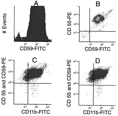
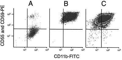
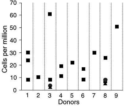
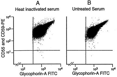
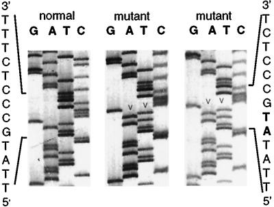
In paroxysmal nocturnal hemoglobinuria (PNH), acquired somatic mutations in the PIG-A gene give rise to clonal populations of red blood cells unable to express proteins linked to the membrane by a glycosylphosphatidylinositol anchor. These proteins include the complement inhibitors CD55 and CD59, and this explains the hypersensitivity to complement of red cells in PNH patients, manifested by intravascular hemolysis. The factors that determine to what extent mutant clones expand have not yet been pinpointed; it has been suggested that existing PNH clones may have a conditional growth advantage depending on some factor (e.g., autoimmune) present in the marrow environment of PNH patients. Using flow cytometric analysis of granulocytes, we now have identified cells that have the PNH phenotype, at an average frequency of 22 per million (range 10-51 per million) in nine normal individuals. These rare cells were collected by flow sorting, and exons 2 and 6 of the PIG-A gene were amplified by nested PCR. We found PIG-A mutations in six cases: four missense, one frameshift, and one nonsense mutation. PNH red blood cells also were identified at a frequency of eight per million. Thus, small clones with PIG-A mutations exist commonly in normal individuals, showing clearly that PIG-A gene mutations are not sufficient for the development of PNH. Because PIG-A encodes an enzyme essential for the expression of a host of surface proteins, the PIG-A gene provides a highly sensitive system for the study of somatic mutations in hematopoietic cells.


Figures

Figure 1
Figure 1
Development of the Methodology. (A) Histogram of normal granulocytes (donor 1) stained with anti-CD59 and goat anti-mouse FITC. A portion (0.17%) of the cells are CD59(−) (fluorescence <101). (B) Two-dimensional scatter analysis of normal granulocytes (donor 1) stained with anti-CD59 and anti-CD55. Here, 0.04% (400 per million)—seen in the lower left quadrant—express neither antigen. (C) Scatter analysis of granulocytes from donor 8, stained with anti-CD55 and anti-CD59, followed by RAM-PE and then CD11b-FITC. A distinct population of PNH cells is seen in the lower right quadrant at a frequency of 7.5 per million and appears similar to the large PNH clone from patient MSK11 (compare with Fig. 2A). (D) Granulocytes from donor 8 stained in parallel to sample in C. The pattern is virtually identical, and the PNH population is present at a frequency of 9.6 per million, confirming the reproducibility of the assay.
Figure 2
Figure 2
(A) Granulocytes from PNH patient MSK11 stained with anti-CD55, anti-CD59, and CD11b-FITC, demonstrating a large PNH clone representing 80% of the cells. (B) Post-sort analysis of collected normal GPI(+) granulocytes from donor 8, demonstrating no residual PNH cells (compare with Fig. 1C). This confirms the ability of the cytometer to accurately sort a rare population. (C) A simulated experiment with PNH cells from patient MSK11 added to sorted GPI(+) cells from donor 1 at a frequency of 25 per million. The PNH population is clearly seen in the lower right quadrant, again confirming the ability of the instrument to identify this rare population. Events, most likely representing debris, also are seen in the lower left quadrant, demonstrating the usefulness of anti-CD11b for excluding these events.
Figure 3
Figure 3
Frequency of PNH granulocytes in nine volunteer donors. Each data point represents the results from a separate blood sample. Three symbols (■, ●, ▴) are used to indicate overlapping data points. PNH cells were identified in all 19 analyses from the donors. The average frequency of PNH cells is 22 per million. The samples from donors 1, 3, 6, and 8 were drawn over a time interval of 6–8 months.
Figure 4
Figure 4
PNH red blood cells in donor 8. Red cells are positively identified by light scatter characteristics and by gating to include glycophorin A(+) events. (A) Flow analysis of red blood cells incubated in a mock Ham test with heat-inactivated serum. PNH cells are clearly seen in the lower right hand quadrant at a frequency of eight per million. (B) A parallel analysis of red blood cells from donor 8 after incubation in the Ham test with untreated serum. The population in the lower right quadrant is reduced by a factor of 6, confirming that these PNH cells exhibit complement sensitivity.
Figure 5
Figure 5
Insertion 196AT in PNH granulocytes from donor 4. Sequence analysis of the noncoding strand is shown for one normal and two representative mutant M13 clones. The insertion/duplication at position 196 (arrow heads, bold type) results in a frameshift and premature chain termination.

References

    1. Oni S-B, Osunkoya B-O, Luzzatto L. Blood. 1970;36:145–152. - PubMed
    1. Ham J-T, Dingle J-H. J Clin Invest. 1939;80:7–12. - PMC - PubMed
    1. Rosse W-F, Ware R-E. Blood. 1995;86:3277–3286. - PubMed
    1. Terstappen L-W, Nguyen M, Huang S, Lazarus H, Medof M-E. Br J Hematol. 1993;84:504–514. - PubMed
    1. Miyata T, Yamada N, Iida Y, Nishimura J, Takeda J, Kitani T, Kinoshita T. N Engl J Med. 1994;330:249–255. - PubMed

Publication types

Substances