. 1999 Apr 27;96(9):5221-6.
doi: 10.1073/pnas.96.9.5221.

Intranuclear delivery of an antiviral peptide mediated by the B subunit of Escherichia coli heat-labile enterotoxin

Affiliations

Intranuclear delivery of an antiviral peptide mediated by the B subunit of Escherichia coli heat-labile enterotoxin

A Loregian et al. Proc Natl Acad Sci U S A. .

Abstract

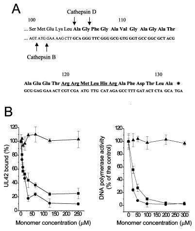
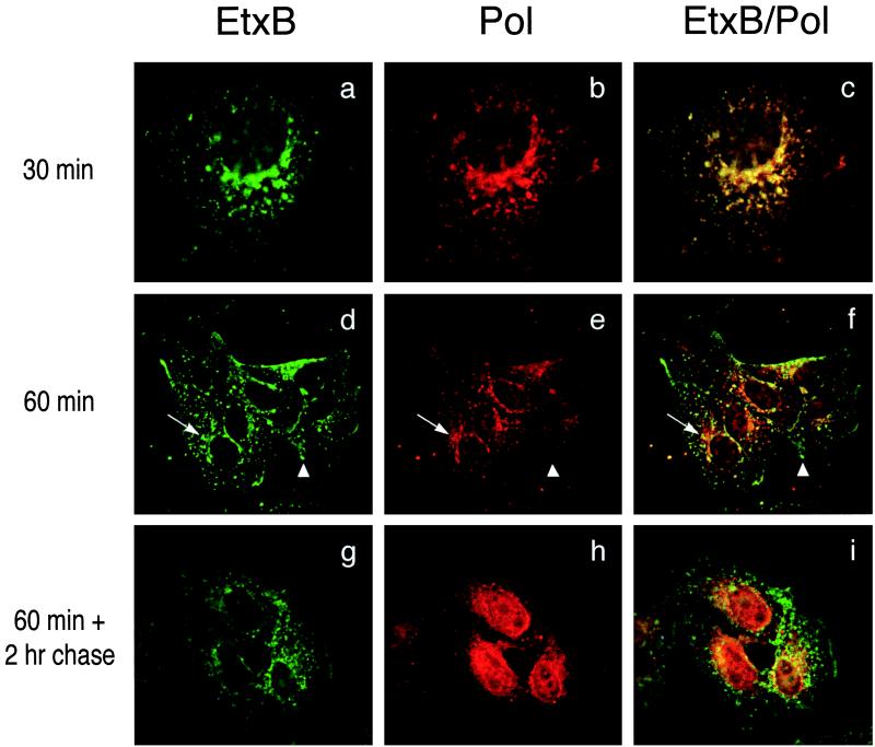
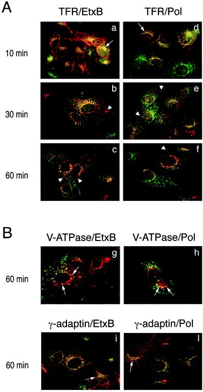
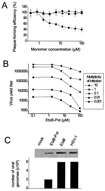
We report an intracellular peptide delivery system capable of targeting specific cellular compartments. In the model system we constructed a chimeric protein consisting of the nontoxic B subunit of Escherichia coli heat-labile enterotoxin (EtxB) fused to a 27-mer peptide derived from the DNA polymerase of herpes simplex virus 1. Viral DNA synthesis takes places in the nucleus and requires the interaction with an accessory factor, UL42, encoded by the virus. The peptide, designated Pol, is able to dissociate this interaction. The chimeric protein, EtxB-Pol, retained the functional properties of both EtxB and peptide components and was shown to inhibit viral DNA polymerase activity in vitro via disruption of the polymerase-UL42 complex. When added to virally infected cells, EtxB-Pol had no effect on adenovirus replication but specifically interfered with herpes simplex virus 1 replication. Further studies showed that the antiviral peptide localized in the nucleus, whereas the EtxB component remained associated with vesicular compartments. The results indicate that the chimeric protein entered through endosomal acidic compartments and that the Pol peptide was cleaved from the chimeric protein before being translocated into the nucleus. The system we describe is suitable for delivery of peptides that specifically disrupt protein-protein interactions and may be developed to target specific cellular compartments.


Figures

Figure 1
Figure 1
Sequence and in vitro activity of the EtxB-Pol chimera. (A) Sequence of the C-terminal 32 residues of the fusion protein is reported, with the amino acids numbered from the first residue of mature EtxB. Boldface letters indicate the region coding the attached Pol peptide. Putative endoprotease cleavage sites are indicated by an arrow; a putative nuclear localization signal is also indicated (underlined sequence). (B) Effect of EtxB (▴), EtxB-Pol (●), and Pol peptide (■) on the POL/UL42 physical interaction as measured by the ELISA interaction assay (Left) and on incorporation of [3H]dTTP into a poly(dA)⋅oligo(dT) template by POL in the presence of UL42 (Right). The graphs show the average of data from three independent experiments together with the SD.
Figure 2
Figure 2
Pol peptide is internalized by EtxB-Pol fusion protein and localizes into the cell nucleus. Vero cells were treated with EtxB-Pol for 30 min (ac) or 1 hr (df) and fixed immediately. Alternatively, cells were treated with the fusion protein for 1 hr, washed, and fixed 2 hr after treatment (gi). All samples then were probed with mAb 118–8, specific for EtxB (EtxB, Left), and with polyclonal antiserum 113, specific for the Pol peptide (Pol, Center) and secondary anti-mouse fluorescein-conjugated or anti-rabbit Texas red-conjugated antibodies, respectively. Images were collected by using a ×100 objective with a deconvolution fluorescence microscope and superimposed (EtxB/Pol, Right). Arrows and arrowheads point at representative EtxB+/Pol+ and EtxB+/Pol− compartments, respectively.
Figure 3
Figure 3
Colocalization of the two EtxB-Pol domains with markers of intracellular compartments. (A) Vero cells were treated with EtxB-Pol for 10, 30, or 60 min and probed either with an antibody specific for TFR and an antibody against EtxB (TFR/EtxB, ac) or with an antibody specific for TFR and the antiserum 113 against the Pol peptide (TFR/Pol, df). Images were analyzed as described in the legend to Fig. 2. Arrows point at an example of TFR+/EtxB+ or TFR+/Pol+ compartments; arrowheads indicate TFR−/EtxB+ or TFR−/Pol+ vesicles. (B) Vero cells were treated with EtxB-Pol for 60 min and probed either with the antibody OSW2 specific for V-ATPase and an antibody specific for EtxB (V-ATPase/EtxB, g), with the antibody OSW2 and the antiserum 113 (V-ATPase/Pol, h), with an antibody specific for γ-adaptin and an antibody against EtxB (γ-adaptin/EtxB, i), or with an antibody specific for γ-adaptin and the antiserum 113 (γ-adaptin/Pol, l). Colocalization of EtxB and Pol moieties of EtxB-Pol with V-ATPase or with γ-adaptin are indicated by arrows.
Figure 4
Figure 4
Effect of different drugs on intranuclear localization of Pol peptide. Vero cells were treated for 1 hr with BFA, bafilomycin A1 (BafA1), or nocodazole (Noc) or not treated (mock). EtxB-Pol was added for 1 hr, and then monolayers were washed and incubated for a further 2 hr in the presence of the inhibitors. Double immunofluorescence was carried out with mAb 118–8 (anti-EtxB, green fluorescence) and rabbit polyclonal serum 113 (anti-Pol peptide, red fluorescence). Images of the same field for both stains were taken with a confocal microscope at a ×100 magnification and superimposed.
Figure 5
Figure 5
The EtxB-Pol fusion protein inhibits HSV-1 replication and DNA synthesis in Vero cells. (A) Inhibition of plaque formation. Confluent Vero cells were infected with HSV-1 at a moi of 0.001 and treated with EtxB-Pol (●), EtxB (▴), or the Pol peptide (■) for 48 hr. The plaque-forming efficiency was calculated as a percentage of plaques compared with an untreated control. The graph shows the mean values and SDs from three independent experiments. (B) Inhibition of virus yield. Vero cells were infected with HSV-1 at different mois and treated with various amounts of EtxB-Pol for 48 hr. Virus progeny was harvested from the infected cells and titered by using a standard plaque assay. (C) Inhibition of viral DNA synthesis. Total DNA was extracted from Vero cells infected with HSV-1 and treated with EtxB-Pol or EtxB or cells that were infected and untreated (HSV-1) or not infected (mock). Samples were digested with BamHI, blotted, and hybridized to a probe complementary to the HSV-1 BamHI Q fragment. Plotted values represent the number of viral genomes relative to autoradiographic bands, quantified by densitometry and correlated with a reference curve as described in the text.

References

    1. Eisenstein E, Schachman H K. In: Protein Function: A Practical Approach. Creighton T E, editor. Oxford: IRL; 1989. pp. 135–175.
    1. Dutia B M, Frame M C, Subak-Sharpe J H, Clark W N, Marsden H S. Nature (London) 1986;321:439–441. - PubMed
    1. Cohen E A, Gaudreau P, Brazeau P, Langelier Y. Nature (London) 1986;321:441–443. - PubMed
    1. Montecucco C, Papini E. Trends Microbiol. 1995;3:165–167. - PubMed
    1. Spangler B D. Microbiol Rev. 1992;56:622–647. - PMC - PubMed

Publication types

MeSH terms

LinkOut - more resources