. 1999 Apr 27;96(9):5268-73.
doi: 10.1073/pnas.96.9.5268.

A neuronal morphologic type unique to humans and great apes

Affiliations

A neuronal morphologic type unique to humans and great apes

E A Nimchinsky et al. Proc Natl Acad Sci U S A. .

Abstract

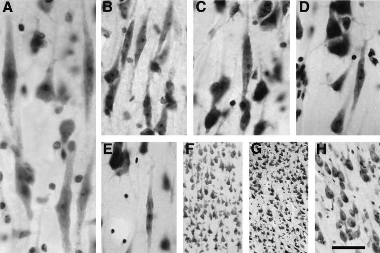
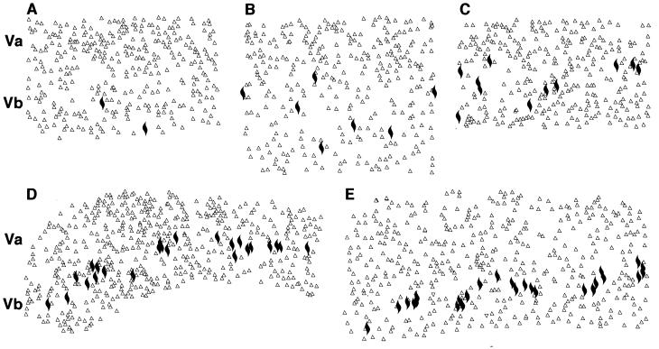
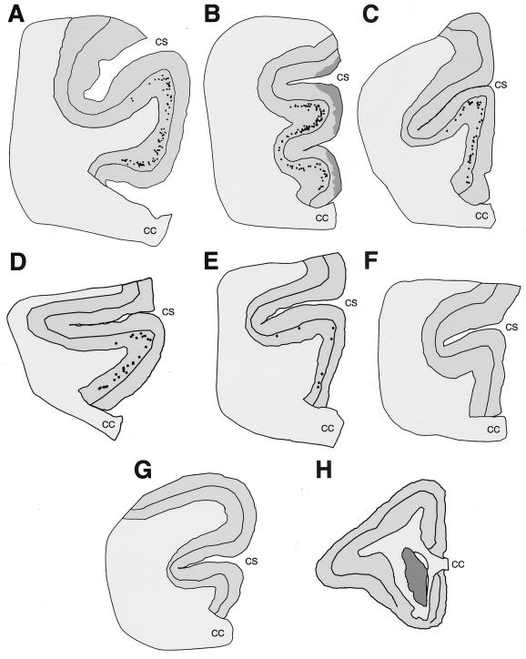
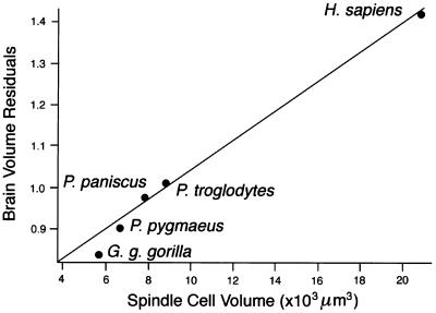
We report the existence and distribution of an unusual type of projection neuron, a large, spindle-shaped cell, in layer Vb of the anterior cingulate cortex of pongids and hominids. These spindle cells were not observed in any other primate species or any other mammalian taxa, and their volume was correlated with brain volume residuals, a measure of encephalization in higher primates. These observations are of particular interest when considering primate neocortical evolution, as they reveal possible adaptive changes and functional modifications over the last 15-20 million years in the anterior cingulate cortex, a region that plays a major role in the regulation of many aspects of autonomic function and of certain cognitive processes. That in humans these unique neurons have been shown previously to be severely affected in the degenerative process of Alzheimer's disease suggests that some of the differential neuronal susceptibility that occurs in the human brain in the course of age-related dementing illnesses may have appeared only recently during primate evolution.


Figures

Figure 1
Figure 1
Morphology of spindle cells in layer Vb of the anterior cingulate cortex in human (A), bonobo (B), common chimpanzee (C), gorilla (D), and orangutan (E). In all of these species the spindle cells display similar morphology and apparent somatic size. Note the clusters of spindle cells in the through-focus photomontage from the human and in the bonobo, whereas isolated neurons are observed in the three other great apes. (F–H) No spindle cells are present in the anterior cingulate cortex of the white-handed gibbon (F), Patas monkey (G), or ring-tailed lemur (H). [Bar = 50 μm (A), 80 μm (B–E and H), and 120 μm (F and G).]
Figure 2
Figure 2
High-magnification computer-generated maps of the localization of spindle neurons in layers V of the anterior cingulate cortex in orangutan (A), gorilla (B), common chimpanzee (C), bonobo (D), and human (E). Spindle cells are represented by solid marks, and the neighboring pyramidal neurons are represented by open triangles. Note the differences in the densities of these neurons among species and the clustered pattern in bonobo and human. AC are 1-mm wide and D and E are 1.5-mm wide.
Figure 3
Figure 3
Computer-generated maps of the distribution of spindle neurons in the anterior cingulate cortex. All maps show a level situated at the genu of the corpus callosum in the human (A), bonobo (B), common chimpanzee (C), gorilla (D), and orangutan (E). The spindle cells are restricted to layer Vb and show much higher numbers in human and the two chimpanzee species, whereas fewer are seen in gorilla and orangutan. No spindle cells are observed in the white-handed gibbon (F), long-tailed macaque monkey (G), and owl monkey (H). The maps are approximately to scale. The darkly shaded areas in B reflect local damage to the superficial layers of the specimen. The caudate nucleus is shaded darkly in H. CC, corpus callosum; CS, cingulate sulcus.
Figure 4
Figure 4
Comparison of spindle cell volume with relative brain volume (see refs. and 20). There is a strong correlation between the volumes of these neurons and brain volume residuals. No such correlation existed for pyramidal and small fusiform neurons of layers V and VI.

References

    1. Frahm H D, Stephan H, Stephan M. J Hirnforsch. 1982;23:375–389. - PubMed
    1. Dunbar R I M. Evol Anthropol. 1998;6:178–190.
    1. Betz W. Zentralbl Med Wiss. 1881;19:193–195. , 209–213, 231–234.
    1. Hammarberg C. Studien über Klinik und Pathologie der Idiotie nebst Untersuchungen über die normale Anatomie des Hirnrinde. Uppsala: Berling; 1895.
    1. Ramón y Cajal S. Textura del Sistema Nervioso del Hombre y de los Vertebrados, Tomo II. Madrid: Nicolás Moya; 1899.

Publication types

LinkOut - more resources