. 1999 Apr 30;284(5415):822-5.
doi: 10.1126/science.284.5415.822.

Chaperonin function: folding by forced unfolding

Affiliations

Chaperonin function: folding by forced unfolding

M Shtilerman et al. Science. .

Abstract

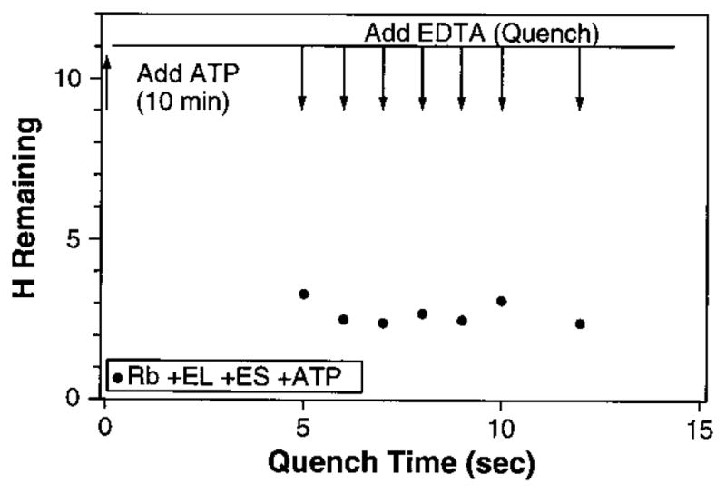
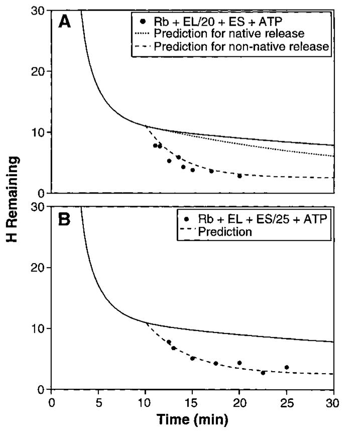
The ability of the GroEL chaperonin to unfold a protein trapped in a misfolded condition was detected and studied by hydrogen exchange. The GroEL-induced unfolding of its substrate protein is only partial, requires the complete chaperonin system, and is accomplished within the 13 seconds required for a single system turnover. The binding of nucleoside triphosphate provides the energy for a single unfolding event; multiple turnovers require adenosine triphosphate hydrolysis. The substrate protein is released on each turnover even if it has not yet refolded to the native state. These results suggest that GroEL helps partly folded but blocked proteins to fold by causing them first to partially unfold. The structure of GroEL seems well suited to generate the nonspecific mechanical stretching force required for forceful protein unfolding.


Figures

Fig. 1
Fig. 1
(Top) The crystal structure of the asymmetric GroEL14-GroES7 complex solved by Xu et al. (5). The two opposed heptameric rings of GroEL are shown in white and yellow. The binding sites for GroES and the substrate protein are in the apical domains between each green and red helix pair (–5, 33). In the less expanded ring (left), which captures the substrate protein, the binding sites are 25 Å from each other. On addition of ATP and GroES, the apical domain of each GroEL subunit twists upward and outward so that the binding sites move apart to a position 33 Å from one another, as shown in the open conformation at the right with the bound GroES removed for clarity. Neighboring binding sites move apart by 8 Å and non-neighboring sites by larger increments, up to 20 Å. A substrate protein tethered to these sites will be forcibly stretched and partially unfolded. [Figure supplied by Z. Xu and P. B. Sigler; see also figure 1 of (32).] (Bottom) A schematic representation of the mechanism of stretch-induced T-H exchange. In the resting state (left) a segment of the substrate protein is tethered between two of the seven peptide binding sites in the apical domain of GroEL. Within the substrate protein a secondary structural element, for example a β-sheet as shown here by the open arrows, protects the radiolabeled amide hydrogens (T) from exchange. During the encapsulation process the rigid body movement of the apical domains causes the peptide binding sites to move further apart (right), generating a stretch-induced unfolding of the substrate protein and rapid exchange of the amide hydrogens (H).
Fig. 2
Fig. 2
Hydrogen-tritium exchange of unfolded RuBisCO. Experimental results (36) monitor the exchange behavior of the well-protected amide hydrogens of unfolded RuBisCO (Rb) when Rb is diluted from denaturing urea into native conditions (pH 8, 22° ± 2°C), where Rb cannot fold without the entire GroEL system. (A) to (C) show the effects on the well-protected hydrogens when the blocked Rb is bound to GroEL (EL) with or without GroES (ES), adenosine diphosphate (ADP), and nucleoside triphosphate [ATP or β,α-imidoadenosine 5′-triphosphate (AMP-PNP)].
Fig. 3
Fig. 3
Single-turnover experiment. ATP was added after 10 min of RuBisCO hydrogen exchange. The upper line shows the number of retained, unexchanged tritiums at that point (from Fig. 2). EDTA was then added at the times shown, before one turnover was completed, leaving the system committed to complete one round of ATP hydrolysis but prohibiting further rounds.
Fig. 4
Fig. 4
Kinetics of substrate protein release. (A) Time-dependent hydrogen exchange in the presence of limiting GroEL (RuBisCO:GroEL: GroES ratio of 1:0.05:1.2). The solid line is from Fig. 2. The upper predicted curve (dotted line) assumes that RuBisCO is released from the complex only when it reaches the native form after an average of 24 turnovers (10), that it loses its carried tritium (except for 2.5 still protected sites) on the first turnover cycle (13 s), and then does not compete for rebinding. The lower predicted curve (dashed line) assumes that each RuBisCO molecule experiences complete tritium loss on one turnover (except for 2.5 protected sites) and is then ejected from the GroEL complex while still unfolded so that it competes with all the other unfolded molecules for rebinding. (B) Time-dependent hydrogen exchange with limiting GroES (RuBisCO: GroEL:GroES = 1:1.2:0.04). The predicted curve assumes that GroES cycles through the RuBisCO-GroEL complexes and induces exchange of the sensitive tritium label on the first RuBisCO turnover. The fitting equation for hydrogen exchange (HX) with non-native protein release on each turnover is H = Aexp[−kb(tto)]exp[−kcat(tto)] + C. The total number of exchangeable hydrogens at the initial 10-min time point (to) is 11, given by 8.5 sensitive hydrogens (A) and 2.5 insensitive ones (C). The background uncatalyzed HX rate during the pertinent time period (10 to 30 min) is kb (0.033 min−1). The chaperone-catalyzed HX rate (kcat) in (A) is 0.23 min−1, given by the 1/20 stoichiometry and 13-s processing time. In (B), kcat is 0.18 min−1, given by the 1/25 stoichiometry and 13-s processing time. The equation for the native protein release curve in (A) was approximated as H = [AN(tto)]exp[−kb(tto)] + C. A, C, and kb are as before. N depends on the linear recovery rate for native protein, expressed in terms of the rate of loss of exchangeable hydrogen label (0.01 min−1), given by the 1/20 stoichiometry, 13-s turnover time, and 24 turnovers per successful RuBisCO folding.

References

    1. Fenton WA, Horwich AL. Protein Sci. 1997;6:743. - PMC - PubMed
    2. Coyle JE, Jaeger J, Groβ M, Robinson CV, Radford SE. Folding Des. 1997;2:R93. - PubMed
    3. Braig K. Curr Opin Struct Biol. 1998;8:159. - PubMed
    1. Sigler PB, et al. Annu Rev Biochem. 1998;67:581. - PubMed
    1. Fenton WA, Kashi Y, Furtak K, Horwich AL. Nature. 1994;371:614. - PubMed
    2. Braig K, et al. :578. ibid.
    1. Roseman AM, Chen S, White H, Braig K, Saibil HR. Cell. 1996;87:241. - PubMed
    1. Xu Z, Horwich AL, Sigler PB. Nature. 1997;388:741. - PubMed

Publication types

MeSH terms