

Review
doi: 10.1128/AAC.43.5.1003.
Aminoglycosides: nephrotoxicity
Affiliations
- PMID: 10223907
- PMCID: PMC89104
- DOI: 10.1128/AAC.43.5.1003
Item in Clipboard
Review
Aminoglycosides: nephrotoxicity
Antimicrob Agents Chemother.
1999 May.
Free PMC article
No abstract available
Figures
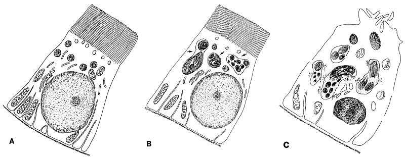
Ultrastructural alterations induced in proximal tubular
cells during aminoglycoside treatment. (A) Control. Changes detected
early on and at low doses (B) consist mainly of the enlargement of
lysosomes, which most likely occurs by fusion of preexisting structures
and which is caused by the progressive deposition of polar lipids which
adopt a concentric lamellar disposition (myelin-like structures, most
commonly referred to as myeloid bodies); the other
subcellular structures are usually well preserved. Later changes or
changes observed with high doses (C) include the apparent rupture of
lysosomes (with the release of myeloid bodies in the cytosol),
extensive mitochondrial swelling and damage, dilatation of the
endoplasmic reticulum cisternae, shedding of the apical brush-border
villi, pericellular membrane discontinuities, and the occurrence of
apoptotic nuclei. These alterations do not necessarily coexist in all
cells. The figure is adapted from reference and
is based on the typical descriptions given in references ,
, , , , , and .
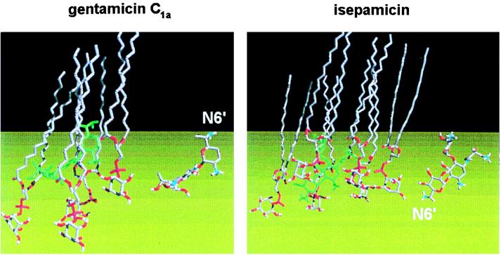
Skeleton views of the mode of assembly of gentamicin
C1a and isepamicin (in green) with phosphatidylinositol
(hydrocarbon is in grey and oxygen and phosphorus atoms are in red to
clearly indicate the polar domain). The two isolated drug molecules,
with the same orientation, are shown on the right for ease of
identification of their various parts (carbons are in grey, oxygens are
in red, and nitrogens are in blue; see Fig. 1 of the companion
minireview [83] for the chemical structures of
isepamicin and gentamicin C1a). The molecular modeling
approach suggests that the orientations and the positions of the two
drugs inserted in the phosphatidylinositol monolayer are entirely
different. First, the N-6′ amino groups are oriented in
opposite directions (toward the lipophilic phase in the case of
gentamicin and toward the water phase for isepamicin. Second,
gentamicin lies above the plane of the inositol moieties and far away
from the water phase (bottom), whereas isepamicin is readily accessible
and is probably therefore more easily displaceable. Similar differences
have been noted between kanamycin A and amikacin (133).
Since both isepamicin and amikacin are characterized by a side chain at
position N-1, these differences have been ascribed to the presence of
this side chain (note that the orientation and position of gentamicin
C1a are very akin to those of gentamicin B, the parent,
unsubstituted compound of isepamicin, and kanamycins). Such changes in
position and orientation are thought to explain the lower inhibitory
potential of amikacin and isepamicin toward the activities of
phospholipases (for discussions, see references ,
, and 131). As outlined in this
minireview and elsewhere (130, 133), inhibition of
phospholipases is probably an important, early event in aminoglycoside
nephrotoxicity. The figure was adapted from reference
, with permission.
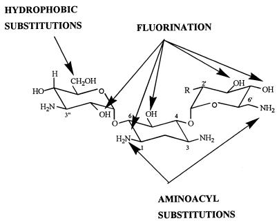
Structural modifications made in kanamycin A (R =
OH) or kanamycin B (R = NH2) (these aminoglycosides
are the most frequently used for chemical modifications) to obtain a
modulation of the potential of the drug to cause lysosomal
phospholipidosis in comparison with the structure of the corresponding
parent compound. The figure is from references , , ,
, , , and .
References
-
- Ademuyiwa O, Ngaha E O, Ubah F O. Vitamin E and selenium in gentamicin nephrotoxicity. Hum Exp Toxicol. 1990;9:281–288. - PubMed
-
- Ali M Z, Goetz M B. A meta-analysis of the relative efficacy and toxicity of single daily dosing versus multiple daily dosing of aminoglycosides. Clin Infect Dis. 1997;24:796–809. - PubMed
-
- Appelkvist E L, Soderstrom M, Nassberger L, Damberg C, Dallner G, DePierre J W. Characterization of the lipid and protein contents of myelin bodies isolated from the renal cortex of gentamicin-treated rats. Biochem Biophys Res Commun. 1991;181:894–901. - PubMed
-
- Assael B M, Chiabrando C, Gagliardi L, Noseda A, Bamonte F, Salmona M. Prostaglandins and aminoglycoside nephrotoxicity. Toxicol Appl Pharmacol. 1985;78:386–394. - PubMed
-
- Aynedjian H S, Nguyen D, Lee H Y, Sablay L B, Bank N. Effects of dietary electrolyte supplementation on gentamicin nephrotoxicity. Am J Med Sci. 1988;295:444–452. - PubMed
Publication types
MeSH terms
Substances
LinkOut - more resources
Full Text Sources
Other Literature Sources
Medical
