

Diversity of substitutions within or adjacent to conserved amino acid motifs of penicillin-binding protein 2X in cephalosporin-resistant Streptococcus pneumoniae isolates
- PMID: 10223944
- PMCID: PMC89251
- DOI: 10.1128/AAC.43.5.1252
Diversity of substitutions within or adjacent to conserved amino acid motifs of penicillin-binding protein 2X in cephalosporin-resistant Streptococcus pneumoniae isolates
Abstract
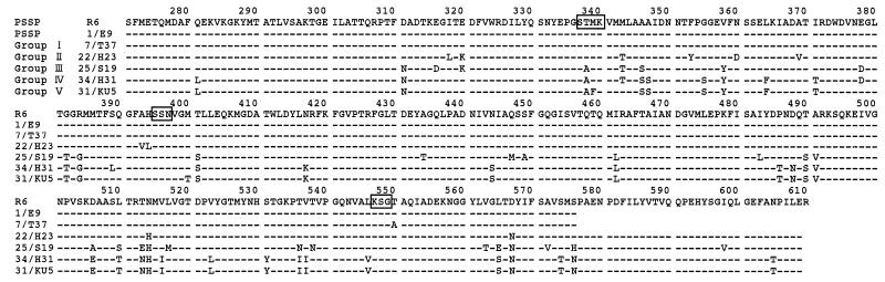
The sequence of an approximately 1.1-kb DNA fragment of the pbp2x gene, which encodes the transpeptidase domain, was determined for 35 clinical isolates of Streptococcus pneumoniae for which the cefotaxime (CTX) MICs varied. Strains with substitutions within a conserved amino acid motif changing STMK to SAFK and a Leu-to-Val change just before the KSG motif were highly resistant to CTX (MIC, >==2 microgram/ml). Strains with substitutions adjacent to SSN or KSG motifs had low-level resistance. The amino acid substitutions were plotted on the three-dimensional crystallographic structure of the transpeptidase domain of PBP2X. Transformants containing pbp2x from strains with high-level CTX resistance increased the CTX MIC from 0. 016 microgram/ml to 0.5 to 1.0 microgram/ml.
Figures

References
-
- Barcus V A, Ghanekar K, Yeo M, Coffey T J, Dowson C G. Genetics of high level penicillin resistance in clinical isolates of Streptococcus pneumoniae. FEMS Microbiol Lett. 1995;126:299–303. - PubMed
-
- Bradley J S, Connor J D. Ceftriaxone failure in meningitis caused by Streptococcus pneumoniae with reduced susceptibility to beta-lactam antibiotics. Pediatr Infect Dis J. 1991;10:871–873. - PubMed
-
- Charlier P, Buisson G, Dideberg O, Wierenga J, Keck W, Laible G, Hakenbeck R. Crystallization of a genetically engineered water-soluble primary penicillin target enzyme. The high molecular mass PBP2x of Streptococcus pneumoniae. J Mol Biol. 1993;232:1007–1009. - PubMed
MeSH terms
Substances
Associated data
- Actions
- Actions
- Actions
- Actions
- Actions
- Actions
- Actions
- Actions
- Actions
LinkOut - more resources
Full Text Sources
Other Literature Sources
