. 1999 May;65(5):2025-31.
doi: 10.1128/AEM.65.5.2025-2031.1999.

Study of the response of a biofilm bacterial community to UV radiation

Affiliations

Study of the response of a biofilm bacterial community to UV radiation

M O Elasri et al. Appl Environ Microbiol. 1999 May.

Abstract

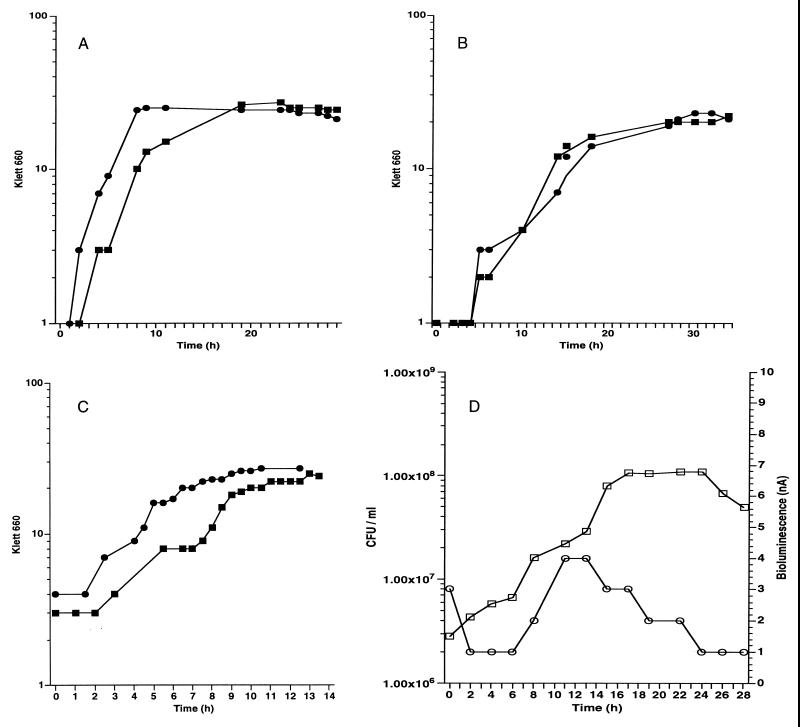
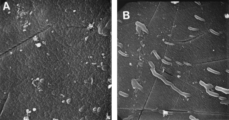
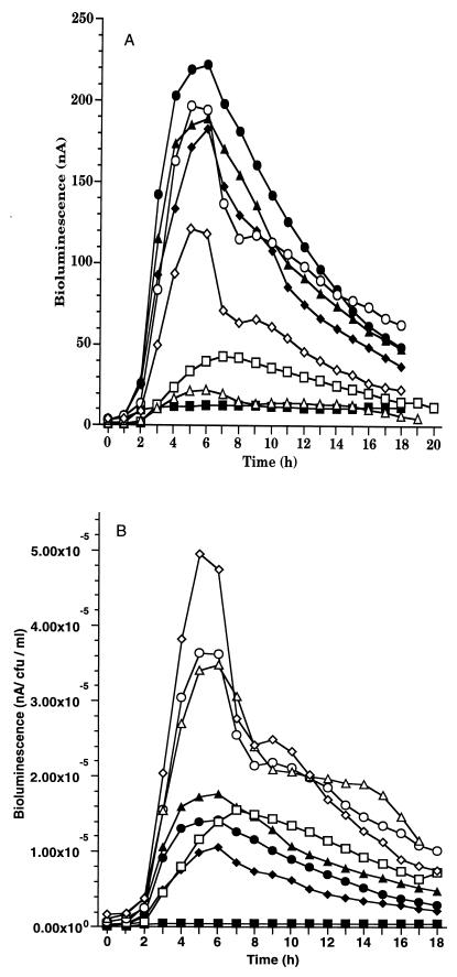
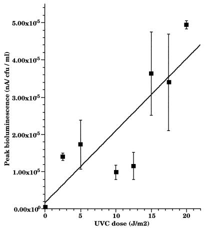
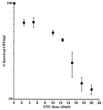
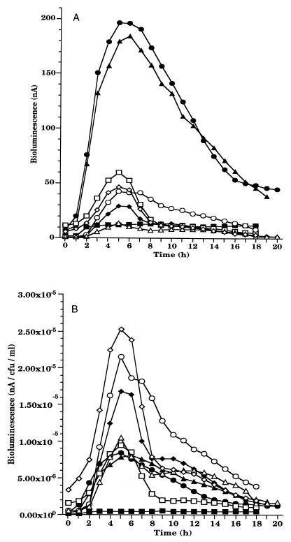
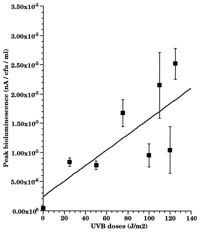
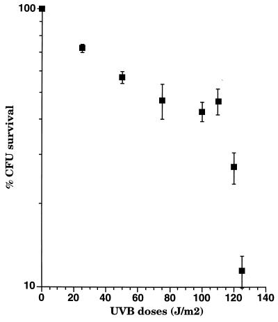
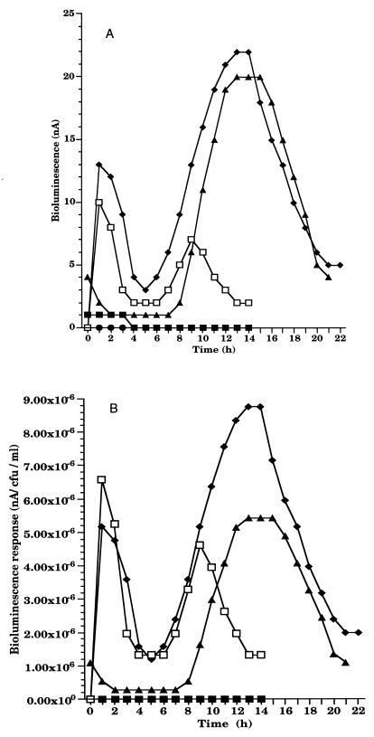
We have developed a bioluminescent whole-cell biosensor that can be incorporated into biofilm ecosystems. RM4440 is a Pseudomonas aeruginosa FRD1 derivative that carries a plasmid-based recA-luxCDABE fusion. We immobilized RM4440 in an alginate matrix to simulate a biofilm, and we studied its response to UV radiation damage. The biofilm showed a protective property by physical shielding against UV C, UV B, and UV A. Absorption of UV light by the alginate matrix translated into a higher survival rate than observed with planktonic cells at similar input fluences. UV A was shown to be effectively blocked by the biofilm matrix and to have no detectable effects on cells contained in the biofilm. However, in the presence of photosensitizers (i.e., psoralen), UV A was effective in inducing light production and cell death. RM4440 has proved to be a useful tool to study microbial communities in a noninvasive manner.


Figures

FIG. 1
FIG. 1
Growth of FRD1 and RM4440. (A) FRD1 in liquid PMM. ■, experiment 1; ●, experiment 2. (B) RM4440 in liquid PMM. ■, experiment 1; ●, experiment 2. (C) RM4440 in liquid PMM supplemented with 1% alginate. ■, experiment 1; ●, experiment 2. (D) RM4440 immobilized in the alginate matrix. Bioluminescence was continuously monitored. □, CFU per milliliter, ○, bioluminescence (nanoamperes). All experiments were done twice.
FIG. 2
FIG. 2
Scanning electron micrographs of the alginate beads containing RM4440 cells. (A) Unirradiated control. (B) Exposure to 10 J of UV C per m2. Stressed cells show filamentation due to inhibition of cell division. Magnification, ca. ×2,000.
FIG. 3
FIG. 3
Bioluminescence response of RM4440 to increasing doses of UV C in biofilm. (A) Total bioluminescence (nanoamperes). (B) Normalized bioluminescence (nanoamperes per CFU per milliliter). UV C doses (J/m2): ■, 0; ●, 2.5; ▴, 5; ⧫, 10; □, 12.5; ○, 15; ▵, 17.5; ◊, 20. Each point represents the mean of three independent experiments.
FIG. 4
FIG. 4
Peak bioluminescence after exposure of RM4440 biofilm to increasing doses of UV C. The peak response was 5 h postirradiation for most doses. The data represent the means of three independent experiments.
FIG. 5
FIG. 5
Survival rate of RM4440 under increasing doses of UV C. Data represent the means of three independent experiments.
FIG. 6
FIG. 6
Bioluminescence response of RM4440 to increasing doses of UV B in biofilm. (A) Total bioluminescence (nanoamperes). (B) Normalized bioluminescence (nanoamperes per CFU per milliliter). UV B doses (J/m2): ■, 0; ●, 25; ▴, 50; ⧫, 75; □, 100; ○, 110; ▵, 120; ◊, 125. Each point represents the mean of three independent experiments.
FIG. 7
FIG. 7
Peak bioluminescence after exposure of RM4440 to increasing doses of UV B in biofilm. The peak response was 5 h postirradiation for most doses. The data represent the means of three independent experiments.
FIG. 8
FIG. 8
Survival rate of RM4440 under increasing doses of UV B. Data represent the means of three independent experiments.
FIG. 9
FIG. 9
Bioluminescence response of RM4440 after exposure to UV A and psoralen. (A) Total bioluminescence (nanoamperes). (B) Normalized bioluminescence (nanoamperes per CFU per milliliter). The biofilm beads were stirred in PMM containing psoralen. ■, UV A only; ●, psoralen only; ▴, ambient light and psoralen; ⧫, 8,000 J of UV A per m2 and psoralen; □, 16,000 J of UV A per m2 and psoralen.

Similar articles

Cited by

References

    1. Averbeck D. Recent advances in psoralen phototoxicity mechanism. Photochem Photobiol. 1989;50:859–882. - PubMed
    1. Bauluz C, Paramio J M, de Vidania R. Further studies on the lethal and mutagenic effects of 8-methoxypsoralen-induced lesions on plasmid DNA. Cell Mol Biol. 1991;37:481–500. - PubMed
    1. Booth M G. Solar ultraviolet radiation and the role of RecA in marine bacteria. Ph.D. dissertation. Stillwater: Oklahoma State University; 1997.
    1. Christensen B E, Characklis W G. Physical and chemical properties of biofilms. In: Characklis W G, Marshall K C, editors. Biofilms. New York, N.Y: John Wiley & Sons, Inc.; 1990. pp. 93–130.
    1. Costerton J W, Cheng K J, Geesey G G, Ladd T I, Nickel J C, Dasgupta M, Marrie T J. Bacterial biofilms in nature and disease. Annu Rev Microbiol. 1987;41:435–464. - PubMed

Publication types

LinkOut - more resources