. 1999 May;65(5):2041-8.
doi: 10.1128/AEM.65.5.2041-2048.1999.

Adhesion of acinetobacter venetianus to diesel fuel droplets studied with In situ electrochemical and molecular probes

Affiliations

Adhesion of acinetobacter venetianus to diesel fuel droplets studied with In situ electrochemical and molecular probes

F Baldi et al. Appl Environ Microbiol. 1999 May.

Abstract

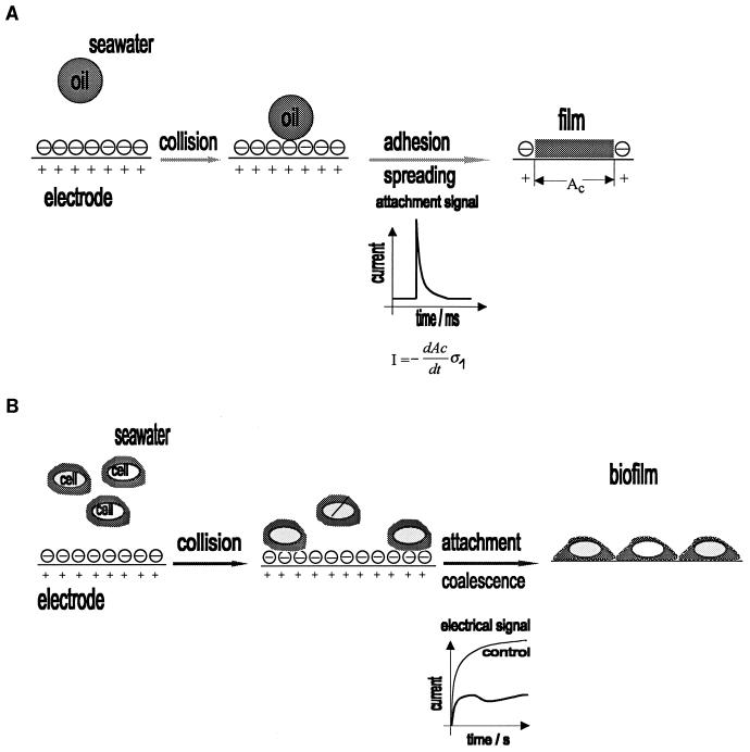
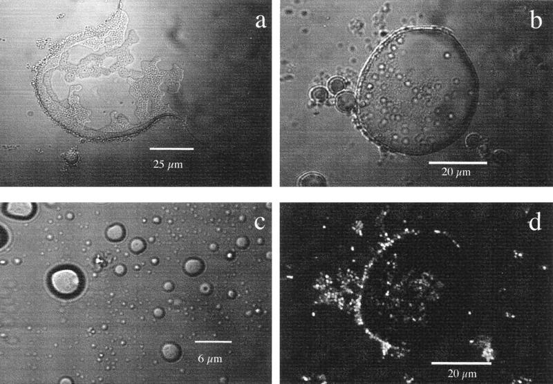
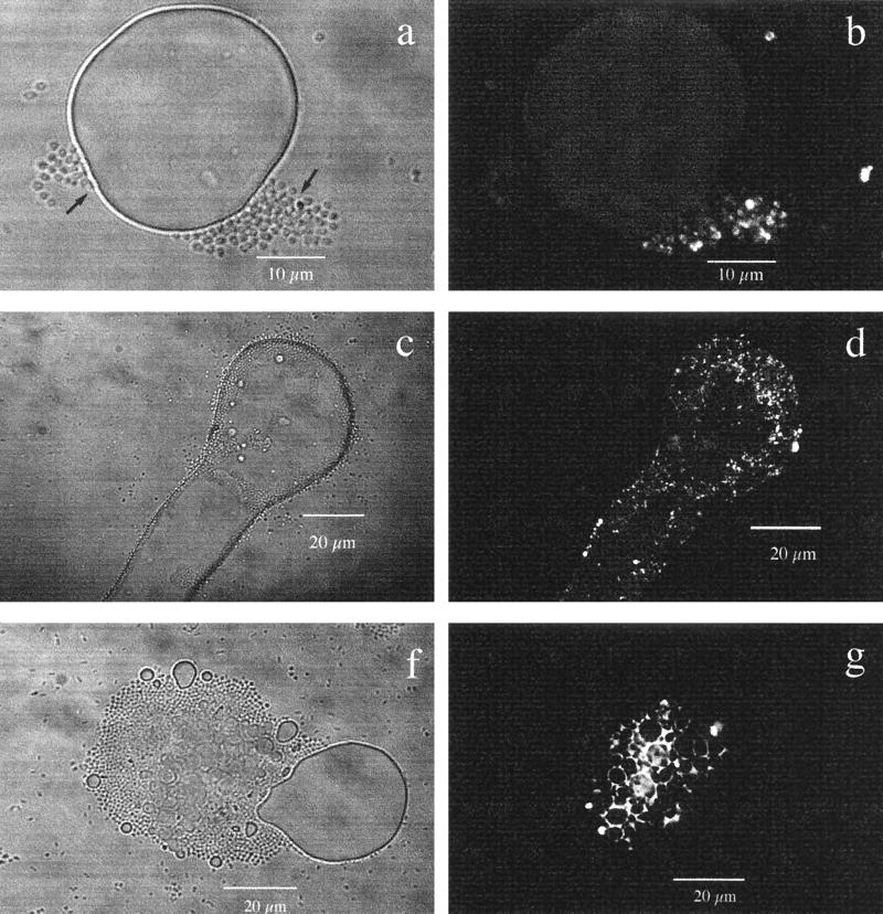
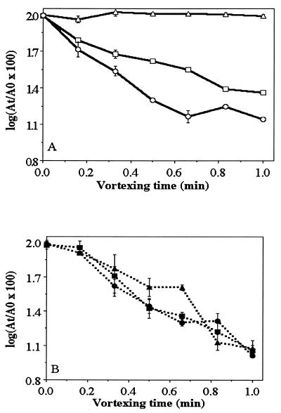
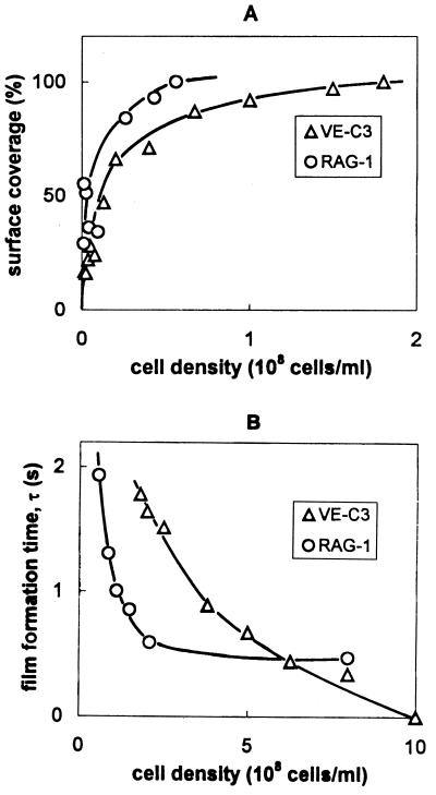
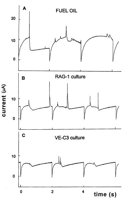
The adhesion of a recently described species, Acinetobacter venetianus VE-C3 (F. Di Cello, M. Pepi, F. Baldi, and R. Fani, Res. Microbiol. 148:237-249, 1997), to diesel fuel (a mixture of C12 to C28 n-alkanes) and n-hexadecane was studied and compared to that of Acinetobacter sp. strain RAG-1, which is known to excrete the emulsifying lipopolysaccharide, emulsan. Oxygen consumption rates, biomass, cell hydrophobicity, electrophoretic mobility, and zeta potential were measured for the two strains. The dropping-mercury electrode (DME) was used as an in situ adhesion sensor. In seawater, RAG-1 was hydrophobic, with an electrophoretic mobility (&mgr;) of -0.38 x 10(-8) m2 V-1 s-1 and zeta potential (zeta) of -4.9 mV, while VE-C3 was hydrophilic, with &mgr; of -0.81 x 10(-8) m2 V-1 s-1 and zeta of -10.5 mV. The microbial adhesion to hydrocarbon (MATH) test showed that RAG-1 was always hydrophobic whereas the hydrophilic VE-C3 strain became hydrophobic only after exposure to n-alkanes. Adhesion of VE-C3 cells to diesel fuel was partly due to the production of capsular polysaccharides (CPS), which were stained with the lectin concanavalin A (ConA) conjugated to fluorescein isothiocyanate and observed in situ by confocal microscopy. The emulsan from RAG-1, which was negative to ConA, was stained with Nile Red fluorochrome instead. Confocal microscope observations at different times showed that VE-C3 underwent two types of adhesion: (i) cell-to-cell interactions, preceding the cell adhesion to the n-alkane, and (ii) incorporation of nanodroplets of n-alkane into the hydrophilic CPS to form a more hydrophobic polysaccharide-n-alkane matrix surrounding the cell wall. The incorporation of n-alkanes as nanodroplets into the CPS of VE-C3 cells might ensure the partitioning of the bulk apolar phase between the aqueous medium and the outer cell membrane and thus sustain a continuous growth rate over a prolonged period.


Figures

FIG. 1
FIG. 1
(A) Adhesion of an oil droplet and its spreading to form a film at the electrode. The electrical signal (transient current) is caused by displacement of the double-layer charge (ς1) from the contact area AC. (B) Adhesion of bacterial cells and formation of molecular contact with the electrode.
FIG. 2
FIG. 2
(A) Oxygen consumption rates determined with Clark’s probe in cultures of Acinetobacter sp. strain RAG-1 (▴) and A. venetianus VE-C3 (▵) grown in mineral medium containing 2 g of diesel fuel liter−1. (B) Protein determination of Acinetobacter sp. strain RAG-1 (▴) and A. venetianus VE-C3 (▵) grown in mineral medium containing 2 g of diesel fuel liter−1.
FIG. 3
FIG. 3
Colonization of diesel fuel by Acinetobacter sp. strain RAG-1. (a) Decrease in surface tension of part of a diesel fuel drop colonized by RAG-1, observed with an optical microscope in the transmission mode after a 3-h incubation at 28°C in mineral medium with diesel fuel (2 g liter−1). (b) Colonization of a diesel fuel droplet (diameter, 56 μm) by RAG-1, showing bacterial adhesion at the rim and on top, observed in the transmission mode after an 8-h incubation at 28°C. (c) Repellent diesel fuel microdroplets of different sizes (from 10 to less than 0.75 μm) dispersed in the medium without attached bacteria after a 24-h incubation. (d) Same image as in panel b but observed by CLSM in the fluorescence mode, formed by 16 overlapped images scanned every 0.8 μm for a total depth of 12.8 μm. This sample was stained with Nile Red fluorochrome for the neutral lipid moiety of emulsan.
FIG. 4
FIG. 4
Colonization of diesel fuel by A. venetianus VE-C3. (a) Light microscopy in the transmission mode, showing aggregates of cells (arrows) before adhesion to the diesel fuel surface after a 6-h incubation at 28°C in mineral medium with 2 g of diesel fuel liter−1. (b) The same image as in panel a but observed by CLSM in the fluorescence mode, obtained by overlapping 20 images scanned every 0.6 μm for a total depth of 12 μm. VE-C3 was stained with ConA-FITC to show polysaccharide residues of glucose and mannose in CPS. Only a fraction of the cells seen in panel a (arrows) have a fluorescent CPS after 6 h of incubation. (c) Transmission mode, showing that the surface tension of a diesel fuel drop colonized by strain VE-C3 decreases and the bacteria at the rim and on top produce elongated and indented shapes after a 12-h incubation. (d) The same image as in panel c but in the fluorescence mode, with the ConA-FITC distribution imaged by CLSM. VE-C3 cells with CPS smear the elongated rim of the diesel fuel drop showing “polysaccharide footprints.” (f) Transmission mode, showing a diesel fuel droplet with a diameter of about 20 μm, with many smaller microdroplets embedded in a microbial aggregate. After a 28-h incubation at 28°C, VE-C3 cells are still attached to diesel fuel droplet. (g) The same image as in panel f but in fluorescence mode with the ConA-FITC distribution imaged by CLSM. The aggregate of cells and diesel fuel microdroplets is glued together by a thick polysaccharide matrix excreted by the bacteria.
FIG. 5
FIG. 5
(A) Cell hydrophobicity of A. venetianus VE-C3 at different times, measured by the MATH test: 0 h (▵) preincubated in PCA medium and then transferred to mineral medium with 2 g of diesel fuel liter−1, then incubated for 12 h (□) and 36 h (○) in mineral medium with 2 g of diesel fuel liter−1. (B) MATH test for Acinetobacter sp. strain RAG-1 under the same conditions as in panel A incubated for 0 h (▴), 12 h (■), and 36 h (●).
FIG. 6
FIG. 6
Adhesion of uninduced VE-C3 and RAG-1 cells at the electrode from their suspensions in 0.1 M NaCl solution. The percentage of surface coverage (A) and the film formation time (B) are plotted as a function of cell density.
FIG. 7
FIG. 7
Electrochemical signals in the diesel fuel-degrading cultures of RAG-1 (curve B) and VE-C3 (curve C) after 3 days of growth. The control (curve A) is an uninoculated dispersion.

References

    1. Baldi F, Minacci A, Saliot A, Mejanelle L, Mozetic̆ P, Turk V, Malej A. Cell lysis and release of particulate polysaccharides in extensive marine mucilage assessed by lipid biomarkers and molecular probes. Mar Ecol Prog Ser. 1997;153:45–57.
    1. Baldi F, Pepi M, Fani R, Di Cello F, Da Ros L, Fossato V U. Complementary degradation of fuel oil in superficial waters and in axenic cultures of aerobic Gram-negative bacteria isolated from Venice Lagoon. Croat Chem Acta. 1997;70:333–346.
    1. Barradas R G, Kimmerle F M. Effect of highly surface-active compounds on polarographic electrode processes. J Electroanal Chem. 1966;11:163–170.
    1. Bonet R, Simon-Pujol M D, Congregado F. Effects of nutrients on exopolysaccharide production and surface properties of Aeromonas salmonicida. Appl Environ Microbiol. 1993;59:2437–2441. - PMC - PubMed
    1. Bradford M M. A rapid and sensitive method for quantitation of microgram quantities of protein utilizing the principle of protein-dye binding. Anal Biochem. 1976;72:248–254. - PubMed

LinkOut - more resources