. 1999 May 3;189(9):1363-72.
doi: 10.1084/jem.189.9.1363.

Inhibition of T cell proliferation by macrophage tryptophan catabolism

Affiliations

Inhibition of T cell proliferation by macrophage tryptophan catabolism

D H Munn et al. J Exp Med. .

Abstract

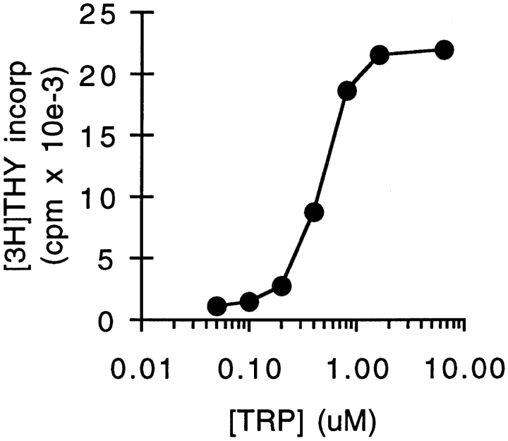
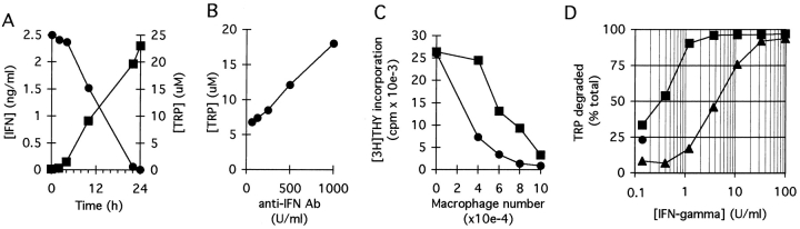
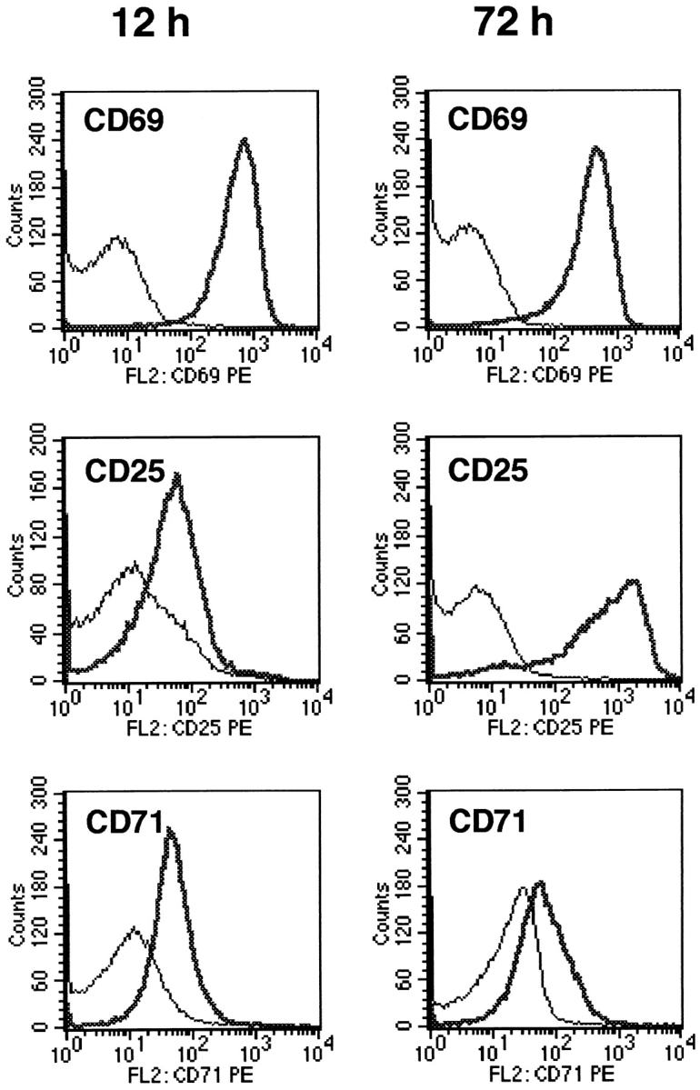
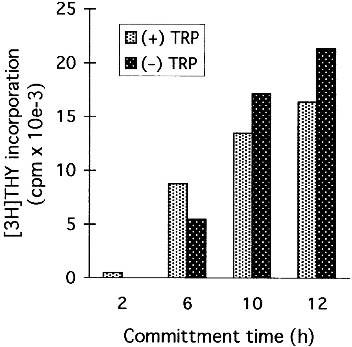
We have recently shown that expression of the enzyme indoleamine 2, 3-dioxygenase (IDO) during murine pregnancy is required to prevent rejection of the allogeneic fetus by maternal T cells. In addition to their role in pregnancy, IDO-expressing cells are widely distributed in primary and secondary lymphoid organs. Here we show that monocytes that have differentiated under the influence of macrophage colony-stimulating factor acquire the ability to suppress T cell proliferation in vitro via rapid and selective degradation of tryptophan by IDO. IDO was induced in macrophages by a synergistic combination of the T cell-derived signals IFN-gamma and CD40-ligand. Inhibition of IDO with the 1-methyl analogue of tryptophan prevented macrophage-mediated suppression. Purified T cells activated under tryptophan-deficient conditions were able to synthesize protein, enter the cell cycle, and progress normally through the initial stages of G1, including upregulation of IL-2 receptor and synthesis of IL-2. However, in the absence of tryptophan, cell cycle progression halted at a mid-G1 arrest point. Restoration of tryptophan to arrested cells was not sufficient to allow further cell cycle progression nor was costimulation via CD28. T cells could exit the arrested state only if a second round of T cell receptor signaling was provided in the presence of tryptophan. These data reveal a novel mechanism by which antigen-presenting cells can regulate T cell activation via tryptophan catabolism. We speculate that expression of IDO by certain antigen presenting cells in vivo allows them to suppress unwanted T cell responses.


Figures

Figure 1
Figure 1
Coculture-conditioned medium is selectively depleted of tryptophan. Human monocytes were allowed to differentiate for 5 d in MCSF. Then, T cells were added and activated with anti-CD3 mAb. Conditioned medium was harvested from cocultures after 48 h and then used to support a second round of activation with fresh T cells. Replicate cultures were supplemented with individual amino acids to the concentrations normally found in RPMI 1640. Control cultures received either fresh medium (CTL) or no supplement (PBS). Proliferation was measured by thymidine incorporation after 72 h.
Figure 2
Figure 2
Dose–response relationship to tryptophan for T cell proliferation. Tryptophan was titrated in coculture-conditioned medium (prepared as described in Fig. 1) and proliferation of T cells measured after 72 h.
Figure 3
Figure 3
Elimination kinetics of tryptophan in cocultures and expression of IDO by MCSF-derived Møs. (A) MCSF-derived Møs were cultured for 24 h with autologous T cells, either with (•) or without (▪) anti-CD3 mAb. The medium was then replaced with fresh medium and supernatant from replicate cultures harvested at the times shown. Tryptophan concentration was assayed spectrofluorometrically as described in Materials and Methods. (B) IFN-γ–inducible IDO mRNA in MCSF-derived Møs. RT-PCR showing IDO expression in MCSF-derived Møs before (lane 4) and after (lanes 1–3) activation for 24 h with recombinant IFN-γ. Starting RNA for the reverse transcriptase reaction in lanes 1–3 was from 20,000, 2000, and 200 activated Møs, respectively, and from 20,000 unactivated Møs in lane 4. Lane 5 shows amplification of human IDO plasmid template giving the expected 182-bp product. (C) HPLC analysis of Mø culture supernatants showing degradation of tryptophan and production of kynurenine. MCSF-derived Møs were preactivated for 24 h with IFN-γ to induce IDO expression, and then the spent medium was replaced 90:10 with fresh medium. Trace 1 shows the analysis of supernatant immediately after adding fresh medium (time 0); trace 2 shows the conditioned medium 24 h later. The number of Møs in these experiments was kept low so that some tryptophan would be detectable at the end of the assay. The traces shown represent the portion of the elution gradient between 28 and 42% acetonitrile (minutes 7.00–10.50), during which kynurenine (K) and tryptophan (T) appeared. The peak labels are positioned at the points at which the purified standards eluted, which were within ±3 s of the corresponding sample peak. Compounds present in culture medium that also absorbed at OD254 (unlabeled peaks) were readily resolved from tryptophan and kynurenine, and the T and K peaks were confirmed by mass spectroscopy (see Materials and Methods). The experiment shown used purified Møs activated with recombinant ligands; identical results were obtained when Møs were activated in coculture with T cells plus mitogen. One of four experiments is shown.
Figure 3
Figure 3
Elimination kinetics of tryptophan in cocultures and expression of IDO by MCSF-derived Møs. (A) MCSF-derived Møs were cultured for 24 h with autologous T cells, either with (•) or without (▪) anti-CD3 mAb. The medium was then replaced with fresh medium and supernatant from replicate cultures harvested at the times shown. Tryptophan concentration was assayed spectrofluorometrically as described in Materials and Methods. (B) IFN-γ–inducible IDO mRNA in MCSF-derived Møs. RT-PCR showing IDO expression in MCSF-derived Møs before (lane 4) and after (lanes 1–3) activation for 24 h with recombinant IFN-γ. Starting RNA for the reverse transcriptase reaction in lanes 1–3 was from 20,000, 2000, and 200 activated Møs, respectively, and from 20,000 unactivated Møs in lane 4. Lane 5 shows amplification of human IDO plasmid template giving the expected 182-bp product. (C) HPLC analysis of Mø culture supernatants showing degradation of tryptophan and production of kynurenine. MCSF-derived Møs were preactivated for 24 h with IFN-γ to induce IDO expression, and then the spent medium was replaced 90:10 with fresh medium. Trace 1 shows the analysis of supernatant immediately after adding fresh medium (time 0); trace 2 shows the conditioned medium 24 h later. The number of Møs in these experiments was kept low so that some tryptophan would be detectable at the end of the assay. The traces shown represent the portion of the elution gradient between 28 and 42% acetonitrile (minutes 7.00–10.50), during which kynurenine (K) and tryptophan (T) appeared. The peak labels are positioned at the points at which the purified standards eluted, which were within ±3 s of the corresponding sample peak. Compounds present in culture medium that also absorbed at OD254 (unlabeled peaks) were readily resolved from tryptophan and kynurenine, and the T and K peaks were confirmed by mass spectroscopy (see Materials and Methods). The experiment shown used purified Møs activated with recombinant ligands; identical results were obtained when Møs were activated in coculture with T cells plus mitogen. One of four experiments is shown.
Figure 4
Figure 4
Inhibition of IDO activity prevents Mø-mediated suppression. (A) 1-methyl-tryptophan inhibits Mø IDO enzyme activity. MCSF-derived Møs were activated with IFN-γ for 24 h to induce IDO expression, and then fresh medium was added as described in Fig. 3, along with 1-methyl-tryptophan (1 mM). Supernatants were analyzed by HPLC for tryptophan (T) and kynurenine (K) immediately after the addition of fresh medium (trace 1) and 24 h later (trace 2). The 1-methyl-tryptophan peak (M) is off scale at the settings used. Control cultures for these experiments (Møs with IFN-γ but without 1-methyl-tryptophan) uniformly had >90% reduction in tryptophan at hour 24, with a corresponding increase in kynurenine, as shown in Fig. 3. The experiment shown used purified Møs activated with recombinant ligands; identical results were obtained when Møs were activated in coculture with T cells plus mitogen. (B) 1-methyl-tryptophan prevents T cell suppression in cocultures. T cells were added to MCSF-derived Møs and activated with anti-CD3 mAb. Replicate cultures were treated with varying concentrations of 1-methyl-tryptophan. Proliferation was measured after 72 h by thymidine incorporation. Controls (○) show proliferation by T cells without Møs at the highest concentration of inhibitor used (there was no effect of inhibitor on T cells alone throughout the range of concentrations shown). (C) A second inhibitor of IDO activity, 6-nitro-tryptophan, showed similar reversal of Mø-mediated inhibition of T cells. Experimental design as in B. (D) Supplementation with high concentrations of tryptophan prevents Mø-mediated suppression. Møs were seeded at low density (CC-lo; 5 × 10⁴ cells/well) and high density (CC-hi; 2 × 10⁵ cells/well) and the medium supplemented with 5× the normal tryptophan concentration. Proliferation was measured at hour 72. Controls show proliferation by Møs alone (M) and T cells alone (T).
Figure 4
Figure 4
Inhibition of IDO activity prevents Mø-mediated suppression. (A) 1-methyl-tryptophan inhibits Mø IDO enzyme activity. MCSF-derived Møs were activated with IFN-γ for 24 h to induce IDO expression, and then fresh medium was added as described in Fig. 3, along with 1-methyl-tryptophan (1 mM). Supernatants were analyzed by HPLC for tryptophan (T) and kynurenine (K) immediately after the addition of fresh medium (trace 1) and 24 h later (trace 2). The 1-methyl-tryptophan peak (M) is off scale at the settings used. Control cultures for these experiments (Møs with IFN-γ but without 1-methyl-tryptophan) uniformly had >90% reduction in tryptophan at hour 24, with a corresponding increase in kynurenine, as shown in Fig. 3. The experiment shown used purified Møs activated with recombinant ligands; identical results were obtained when Møs were activated in coculture with T cells plus mitogen. (B) 1-methyl-tryptophan prevents T cell suppression in cocultures. T cells were added to MCSF-derived Møs and activated with anti-CD3 mAb. Replicate cultures were treated with varying concentrations of 1-methyl-tryptophan. Proliferation was measured after 72 h by thymidine incorporation. Controls (○) show proliferation by T cells without Møs at the highest concentration of inhibitor used (there was no effect of inhibitor on T cells alone throughout the range of concentrations shown). (C) A second inhibitor of IDO activity, 6-nitro-tryptophan, showed similar reversal of Mø-mediated inhibition of T cells. Experimental design as in B. (D) Supplementation with high concentrations of tryptophan prevents Mø-mediated suppression. Møs were seeded at low density (CC-lo; 5 × 10⁴ cells/well) and high density (CC-hi; 2 × 10⁵ cells/well) and the medium supplemented with 5× the normal tryptophan concentration. Proliferation was measured at hour 72. Controls show proliferation by Møs alone (M) and T cells alone (T).
Figure 5
Figure 5
IFN-γ and CD40L act synergistically to induce IDO. (A) MCSF-derived Møs were cocultured with T cells and anti-CD3 mAb. Following lymphocyte addition, culture supernatants were harvested at the times shown and assayed for IFN-γ (▪, left axis) and tryptophan concentration (•, right axis). (B) Møs and T cells were cocultured with mitogen in the presence of various concentrations of neutralizing anti–IFN-γ antiserum. Tryptophan concentration in culture supernatants was determined after 18 h. A low density of Møs was used for these experiments so as not to obscure the effect of IFN-γ. (C) Møs were cultured at a range of seeding densities as shown, and then T cells and anti-CD3 mAb were added either with (▪) or without (•) neutralizing antibodies to IFN-γ (100 neutralizing U/ml). Antibodies to IFN-γ reduced the effectiveness of Møs in suppressing T cells, particularly when the number of Møs was limiting. (D) MCSF-derived Møs were cultured for 24 h with various concentrations of recombinant IFN-γ, either in the presence (▪) or absence (▴) of recombinant CD40L (500 ng/ml). At the end of the activation period, culture supernatants were assayed for the concentration of tryptophan remaining. The single round point shows tryptophan degradation in response to CD40L alone.
Figure 6
Figure 6
T cells do not enter S phase in the absence of tryptophan. T cells were activated with immobilized anti-CD3 mAb plus anti-CD28, either in chemically defined tryptophan-free medium (▪) or in the same medium supplemented with 25 μM tryptophan (•). DNA synthesis was assayed by thymidine incorporation at the times shown.
Figure 7
Figure 7
Expression of activation markers on T cells deprived of tryptophan. T cells were activated in tryptophan-free medium using immobilized anti-CD3/CD28 (heavy trace), or cultured under identical conditions but without anti-CD3/CD28 (light trace). At the times shown, both groups were harvested and stained for expression of activation markers as described in Materials and Methods.
Figure 8
Figure 8
Production of IFN-γ and IL-2 by T cells deprived of tryptophan. T cells were activated with anti-CD3 mAb in tryptophan-free medium (▪) or in the same medium supplemented with 25 μM tryptophan (•) and the concentration of IFN-γ (A) and IL-2 (B) in culture supernatants determined at the times shown.
Figure 9
Figure 9
T cells that have entered the tryptophan-sensitive arrested state retain their position in mid-G1. (A) T cells were activated in tryptophan-free medium using immobilized anti-CD3/CD28 (•). After a period of preactivation (24–72 h with similar results; 48 h in the experiments shown), tryptophan was added and the time to entry into S phase determined (defined as the initiation of thymidine incorporation). Replicate aliquots of cells were activated in tryptophan-containing medium without the 48-h preincubation period (▪). Lag time in each case was defined as the time to initiation of S phase from the point at which cells saw both tryptophan and anti-CD3. The arrow shows that the lag time to S phase was shortened by 12–16 h due to preactivation in the absence of tryptophan, suggesting that this portion of G1 had been accomplished before the point at which cells arrested. Representative of  seven experiments at 36, 48, and 72 h, all showing the same lag time to S phase. (B) T cells were activated with anti-CD3/CD28 in the presence (▪) or absence (•) of tryptophan. After 14 h (the time of the putative arrest point estimated from A), tryptophan was added to the tryptophan-deficient cultures and entry into S phase determined. T cells rescued at hour 14 showed no delay compared with controls.
Figure 10
Figure 10
T cells undergo normal commitment to TCR-independent activation in the absence of tryptophan. T cells were exposed to immobilized anti-CD3/CD28 for 2–12 h in the presence (light bars) or absence (dark bars) of tryptophan. At the times shown, cells were removed from contact with anti-CD3. After transfer, tryptophan was added to the tryptophan-deficient cultures, and all groups were continued out to hour 48. Cells were transferred in their own conditioned medium without washing and continued to receive anti-CD28 throughout. At hour 48, all groups were assayed for proliferation by thymidine incorporation. The 2-h time point (no proliferation after transfer) is included as a control to confirm that there was no carryover of anti-CD3 into the new cultures.
Figure 11
Figure 11
T cells require TCR signaling to exit the arrested state. T cells were activated for 48 h in the absence of tryptophan using immobilized anti-CD3/CD28. To simulate loss of contact with the APC, T cells were removed from the immobilized anti-CD3, washed, and returned to culture in medium containing 25 μm tryptophan. Upon replating, replicate cultures received immobilized anti-CD3, anti-CD28, or both.

References

    1. Fazekas de St. Groth B. The evolution of self-tolerance: a new cell arises to meet the challenge of self-reactivity. Immunol Today. 1998;19:448–454. - PubMed
    1. Banchereau J, Steinman RM. Dendritic cells and the control of immunity. Nature. 1998;392:245–252. - PubMed
    1. Munn DH, Armstrong E. Cytokine regulation of human monocyte differentiation in vitro: the tumor-cytotoxic phenotype induced by macrophage colony-stimulating factor is developmentally regulated by interferon γ. Cancer Res. 1993;53:2603–2613. - PubMed
    1. Munn DH, Pressey J, Beall AC, Hudes R, Alderson MR. Selective activation-induced apoptosis of peripheral T cells imposed by macrophages: a potential mechanism of antigen-specific peripheral lymphocyte deletion. J Immunol. 1996;156:523–532. - PubMed
    1. Shimizu T, Nomiyama S, Hirata F, Hayaishi O. Indoleamine 2,3-dioxygenase: purification and some properties. J Biol Chem. 1978;253:4700–4706. - PubMed

Publication types

MeSH terms