. 1999 May 3;189(9):1383-90.
doi: 10.1084/jem.189.9.1383.

Grf40, A novel Grb2 family member, is involved in T cell signaling through interaction with SLP-76 and LAT

Affiliations

Grf40, A novel Grb2 family member, is involved in T cell signaling through interaction with SLP-76 and LAT

H Asada et al. J Exp Med. .

Abstract

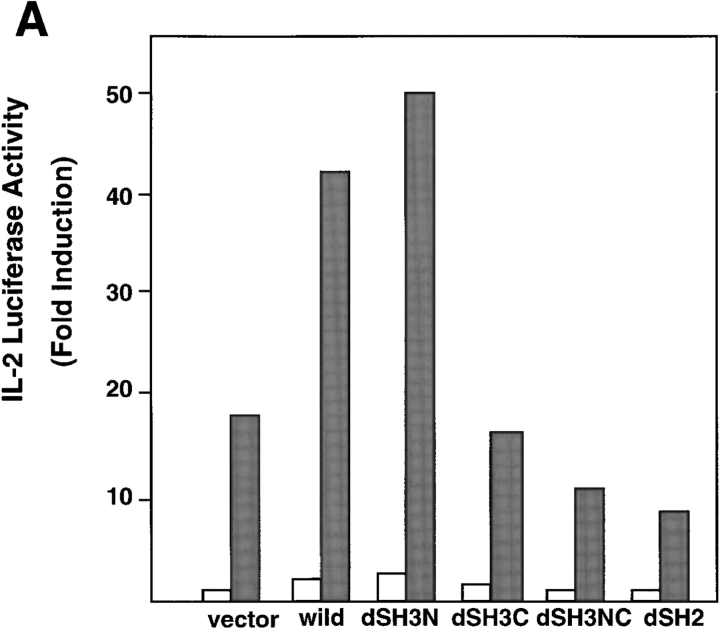
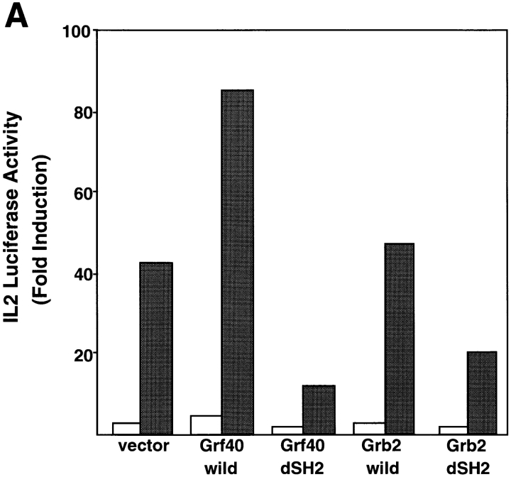
We molecularly cloned a new Grb2 family member, named Grf40, containing the common SH3-SH2-SH3 motif. Expression of Grf40 is predominant in hematopoietic cells, particularly T cells. Grf40 binds to the SH2 domain-containing leukocyte protein of 76 kD (SLP-76) via its SH3 domain more tightly than Grb2. Incidentally, Grf40 binds to linker for activation of T cells (LAT) possibly via its SH2 domain. Overexpression of wild-type Grf40 in Jurkat cells induced a significant increase of SLP-76-dependent interleukin (IL)-2 promoter and nuclear factor of activated T cell (NF-AT) activation upon T cell receptor (TCR) stimulation, whereas the COOH-terminal SH3-deleted Grf40 mutant lacked any recognizable increase in IL-2 promoter activity. Furthermore, the SH2-deleted Grf40 mutant led to a marked inhibition of these regulatory activities, the effect of which is apparently stronger than that of the SH2-deleted Grb2 mutant. Our data suggest that Grf40 is an adaptor molecule involved in TCR-mediated signaling through a more efficient interaction than Grb2 with SLP-76 and LAT.


Figures

Figure 1
Figure 1
Schematic structure and expression of Grf40. (A) The schematic structure of Grf40 was compared with Grb2 and Grap. The percentages of amino acid identity of the SH3 and SH2 domains of Grf40 with Grb2 and Grap were indicated in terms of their SH3 and SH2 domains. (B) 107 cells of a variety of human cell lines were immunoprecipitated (IP) and immunoblotted (IB) with anti-Grf40, anti-Grb2, or preimmune (control [Cont]) serum. (C) Northern blot analyses were performed with the various human tissues indicated. The blots were first hybridized with the Grf40 probe, then rehybridized with β-actin probe.
Figure 2
Figure 2
Coimmunoprecipitation of SLP-76 and LAT with Grf40 in Jurkat cells. Jurkat cells were stimulated with (+) or without (−) OKT3 for 3 min, and their lysates were immunoprecipitated (IP) with anti-Grf40 or preimmune (control) serum. The immunoprecipitates were separated by SDS-PAGE and then immunoblotted (IB) with antiphosphotyrosine mAbs (A), and with anti–SLP-76 antiserum or anti-LAT Ab (B). COS7 cells were transiently transfected with 10 μg expression plasmids for Myc-tagged wild-type Grf40 (wild) or four Myc-tagged Grf40 mutants (dSH3C, dSH2, dSH3N, and dSH3NC), together with 10 μg expression plasmids for SLP-76 by electroporation, and then incubated for 48 h. Their lysates were immunoprecipitated with anti–SLP-76 or preimmune (control) serum, and then immunoblotted with anti-Myc mAb or anti–SLP-76 antiserum (C). COS7 cells were transiently transfected with 10 μg expression plasmids for Flag-tagged wild-type SLP-76 (wild), four Flag-tagged SLP-76 mutants (157-533, 217-533, 241-533, and 281-533), or an empty vector (control), together with 10 μg expression plasmids for Myc-tagged Grf40 by electroporation, and then incubated for 48 h. Their lysates were immunoprecipitated with anti-Flag mAb, and then immunoblotted with anti-Myc or anti-Flag mAb (D).
Figure 3
Figure 3
Competitive binding ability of Grf40 and Grb2 to SLP-76. COS7 cells were transiently transfected with the indicated doses of pFlagSLP (SLP-76), 2.5 μg of pMycGrf40 (Grf40), and 2.5 μg of pMycGrb2 (Grb2) (A), or 0.2 μg of pFlagSLP, 2.5 μg of pMycGrf40, and the indicated doses of pMycGrb2 (B). The cells were incubated for 48 h, and their lysates were immunoprecipitated (IP) with anti-Flag mAb and then immunoblotted (IB) with anti-Myc polyclonal Ab or anti-Flag mAb (top and middle). The expression levels of Myc-tagged Grf40 and Myc-tagged Grb2 were quantified by immunoblotting with anti-Myc mAb (bottom).
Figure 4
Figure 4
Effects of Grf40 mutants on TCR-mediated activation of IL-2 promoter and NF-AT activities. Jurkat cells were transfected with 10 μg of IL-2-Luc (A) or 10 μg of NF-AT-Luc (B), along with 10 μg of SLP-76 and 10 μg of an empty vector (vector), Myc-tagged wild-type Grf40 (wild), or various Grf40 mutants (dSH3N, dSH3C, dSH3NC, and dSH2). The cells were left unstimulated (white bars) or were stimulated (black bars) with 10 μg/ml OKT3 plus 50 ng/ml PMA, and assayed for luciferase activity. Results are shown as fold induction of luciferase activity compared with the activity in unstimulated cells transfected with the empty vector (∼5,000 relative light units [RLU]). These results are representative of three comparable experiments.
Figure 4
Figure 4
Effects of Grf40 mutants on TCR-mediated activation of IL-2 promoter and NF-AT activities. Jurkat cells were transfected with 10 μg of IL-2-Luc (A) or 10 μg of NF-AT-Luc (B), along with 10 μg of SLP-76 and 10 μg of an empty vector (vector), Myc-tagged wild-type Grf40 (wild), or various Grf40 mutants (dSH3N, dSH3C, dSH3NC, and dSH2). The cells were left unstimulated (white bars) or were stimulated (black bars) with 10 μg/ml OKT3 plus 50 ng/ml PMA, and assayed for luciferase activity. Results are shown as fold induction of luciferase activity compared with the activity in unstimulated cells transfected with the empty vector (∼5,000 relative light units [RLU]). These results are representative of three comparable experiments.
Figure 5
Figure 5
Comparison of blocking effects between Grf40-dSH2 and Grb2-dSH2 on TCR-mediated stimulation of IL-2 promoter activity. (A) Jurkat cells were transfected with 10 μg of IL-2-Luc along with 10 μg of SLP-76 and 10 μg of an empty vector (vector), Myc-tagged Grf40 wild-type, Grf40-dSH2, Grb2 wild-type, or Grb2-dSH2. The cells were left unstimulated (white bars) or were stimulated (black bars) with OKT3 plus PMA. (B) Jurkat cells were transfected with 10 μg of IL-2-Luc along with 10 μg of SLP-76 and the indicated doses of Grf40-dSH2 (squares) or Grb2-dSH2 (circles). The cells were left unstimulated (open symbols) or were stimulated (filled symbols) with OKT3 plus PMA, and assayed for luciferase activity. Luciferase activity was indicated as average fold induction ± SE from three independent experiments compared with unstimulated cells transfected with the empty vector (∼5,000 RLU).
Figure 5
Figure 5
Comparison of blocking effects between Grf40-dSH2 and Grb2-dSH2 on TCR-mediated stimulation of IL-2 promoter activity. (A) Jurkat cells were transfected with 10 μg of IL-2-Luc along with 10 μg of SLP-76 and 10 μg of an empty vector (vector), Myc-tagged Grf40 wild-type, Grf40-dSH2, Grb2 wild-type, or Grb2-dSH2. The cells were left unstimulated (white bars) or were stimulated (black bars) with OKT3 plus PMA. (B) Jurkat cells were transfected with 10 μg of IL-2-Luc along with 10 μg of SLP-76 and the indicated doses of Grf40-dSH2 (squares) or Grb2-dSH2 (circles). The cells were left unstimulated (open symbols) or were stimulated (filled symbols) with OKT3 plus PMA, and assayed for luciferase activity. Luciferase activity was indicated as average fold induction ± SE from three independent experiments compared with unstimulated cells transfected with the empty vector (∼5,000 RLU).

References

    1. Weiss A, Littman DR. Signal transduction by lymphocyte antigen receptors. Cell. 1994;76:263–274. - PubMed
    1. Cantrell D. T cell antigen receptor signal transduction pathways. Annu Rev Immunol. 1996;14:259–274. - PubMed
    1. Wange RL, Samelson LE. Complex complexes: signaling at the TCR. Immunity. 1996;5:197–205. - PubMed
    1. Koretzky GA. The role of Grb2-associated proteins in T-cell activation. Immunol Today. 1997;18:401–406. - PubMed
    1. Sieh M, Batzer A, Schlessinger J, Weiss A. GRB2 and phospholipase C-gamma 1 associate with a 36- to 38-kilodalton phosphotyrosine protein after T-cell receptor stimulation. Mol Cell Biol. 1994;14:4435–4442. - PMC - PubMed

Publication types

MeSH terms

Associated data