. 1999 May 3;189(9):1399-412.
doi: 10.1084/jem.189.9.1399.

The P190, P210, and P230 forms of the BCR/ABL oncogene induce a similar chronic myeloid leukemia-like syndrome in mice but have different lymphoid leukemogenic activity

Affiliations

The P190, P210, and P230 forms of the BCR/ABL oncogene induce a similar chronic myeloid leukemia-like syndrome in mice but have different lymphoid leukemogenic activity

S Li et al. J Exp Med. .

Abstract

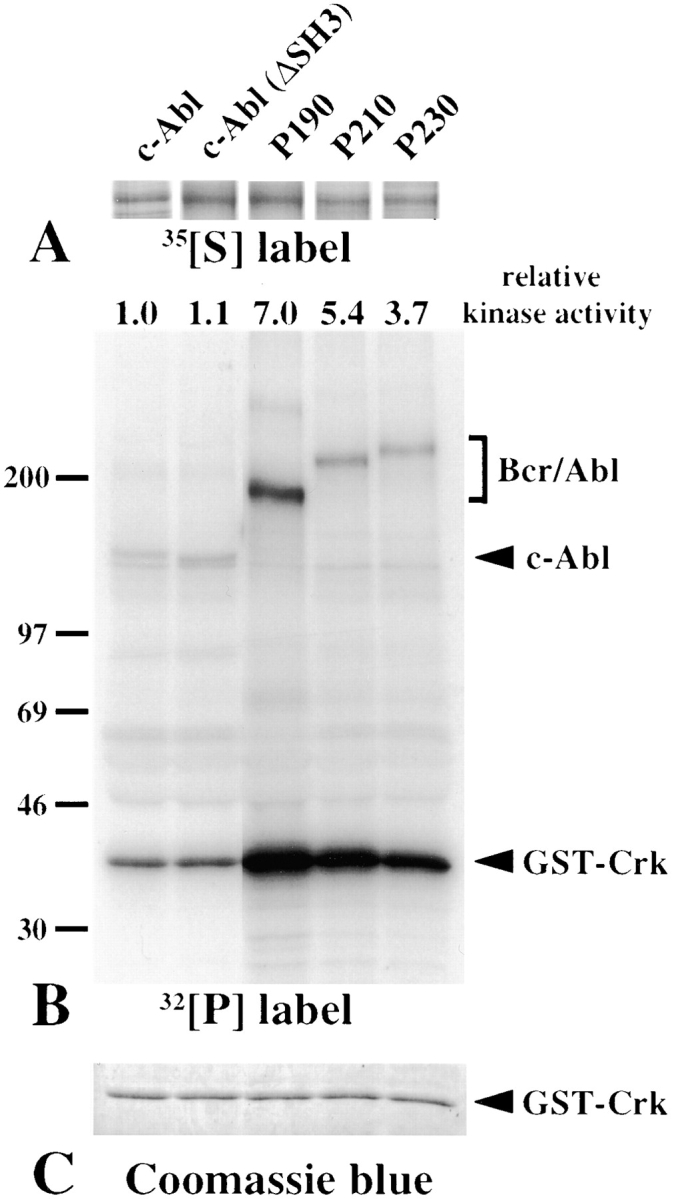
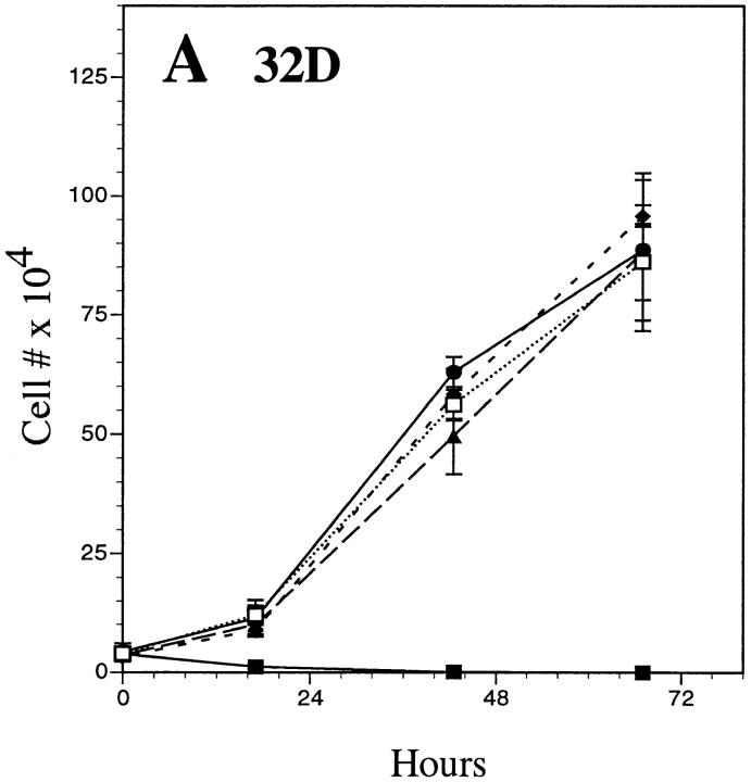
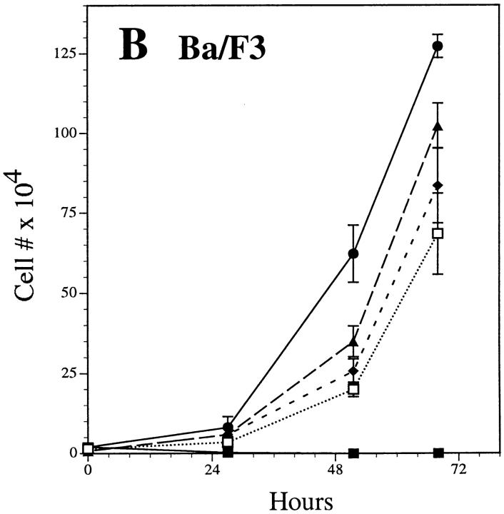
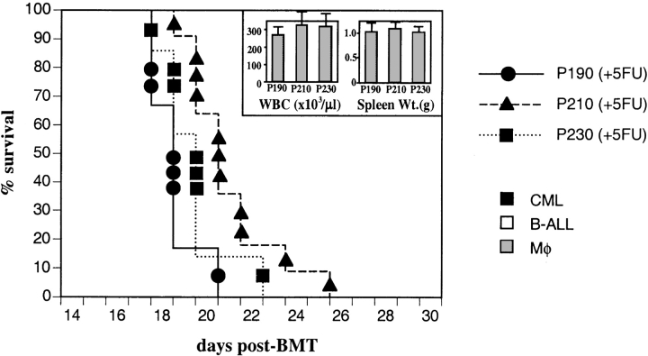
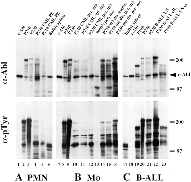
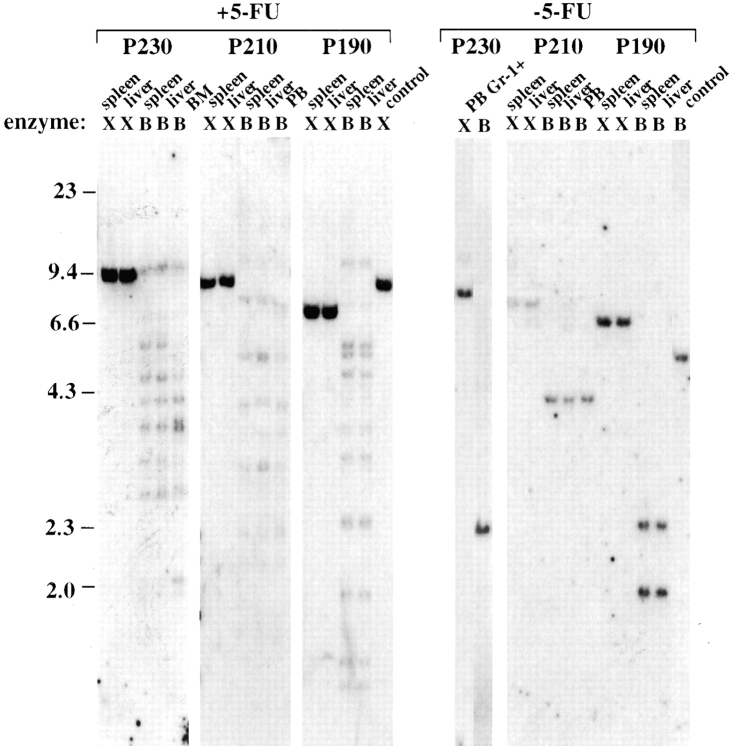
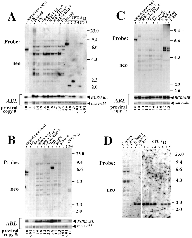
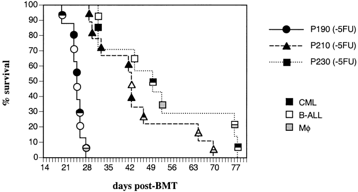
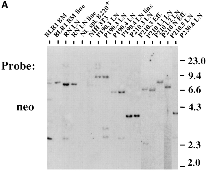
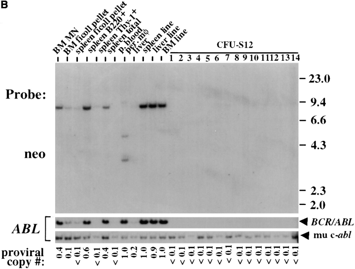
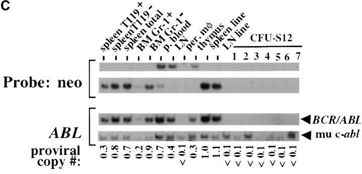
The product of the Philadelphia chromosome (Ph) translocation, the BCR/ABL oncogene, exists in three principal forms (P190, P210, and P230 BCR/ABL) that are found in distinct forms of Ph-positive leukemia, suggesting the three proteins have different leukemogenic activity. We have directly compared the tyrosine kinase activity, in vitro transformation properties, and in vivo leukemogenic activity of the P190, P210, and P230 forms of BCR/ABL. P230 exhibited lower intrinsic tyrosine kinase activity than P210 and P190. Although all three oncogenes transformed both myeloid (32D cl3) and lymphoid (Ba/F3) interleukin (IL)-3-dependent cell lines to become independent of IL-3 for survival and growth, their ability to stimulate proliferation of Ba/F3 lymphoid cells differed and correlated directly with tyrosine kinase activity. In a murine bone marrow transduction/transplantation model, the three forms of BCR/ABL were equally potent in the induction of a chronic myeloid leukemia (CML)-like myeloproliferative syndrome in recipient mice when 5-fluorouracil (5-FU)-treated donors were used. Analysis of proviral integration showed the CML-like disease to be polyclonal and to involve multiple myeloid and B lymphoid lineages, implicating a primitive multipotential target cell. Secondary transplantation revealed that only certain minor clones gave rise to day 12 spleen colonies and induced disease in secondary recipients, suggesting heterogeneity among the target cell population. In contrast, when marrow from non- 5-FU-treated donors was used, a mixture of CML-like disease, B lymphoid acute leukemia, and macrophage tumors was observed in recipients. P190 BCR/ABL induced lymphoid leukemia with shorter latency than P210 or P230. The lymphoid leukemias and macrophage tumors had provirus integration patterns that were oligo- or monoclonal and limited to the tumor cells, suggesting a lineage-restricted target cell with a requirement for additional events in addition to BCR/ABL transduction for full malignant transformation. These results do not support the hypothesis that P230 BCR/ABL induces a distinct and less aggressive form of CML in humans, and suggest that the rarity of P190 BCR/ABL in human CML may reflect infrequent BCR intron 1 breakpoints during the genesis of the Ph chromosome in stem cells, rather than intrinsic differences in myeloid leukemogenicity between P190 and P210.


Figures

Figure 1
Figure 1
In vitro kinase activity of Bcr/Abl proteins. The indicated Abl proteins were expressed by transient transfection of 293 cells, immunoprecipitated, and incubated with [γ-32P]ATP and GST-Crk substrate as described in Materials and Methods. (A) 35S incorporation into immunoprecipitated Abl proteins. (B) 32P label. The positions of c-Abl and Bcr/Abl proteins and the GST-Crk substrate are indicated. (C) Coomassie blue stain indicating equal amounts of GST-Crk substrate in each reaction. The transkinase activity of each Abl protein, relative to c-Abl, is given at the top.
Figure 2
Figure 2
Proliferation of factor-dependent hematopoietic cells transformed by the three BCR/ ABL oncogenes. (A) 32D cl3 myeloid cells. (B) Ba/F3 lymphoid cells. In each case, MSCVneo vector–transduced cells were grown in either the presence or absence of IL-3. The difference between the day 2 mean cell number for Ba/F3-MSCVneo (+IL-3) and Ba/F3-P190, and between Ba/F3-P190 and Ba/F3-P210 or Ba/F3-P230 was significant (P = 0.05, t test). The difference between the day 2 mean cell number between Ba/F3-P210 and Ba/F3-P230 was not significant.
Figure 2
Figure 2
Proliferation of factor-dependent hematopoietic cells transformed by the three BCR/ ABL oncogenes. (A) 32D cl3 myeloid cells. (B) Ba/F3 lymphoid cells. In each case, MSCVneo vector–transduced cells were grown in either the presence or absence of IL-3. The difference between the day 2 mean cell number for Ba/F3-MSCVneo (+IL-3) and Ba/F3-P190, and between Ba/F3-P190 and Ba/F3-P210 or Ba/F3-P230 was significant (P = 0.05, t test). The difference between the day 2 mean cell number between Ba/F3-P210 and Ba/F3-P230 was not significant.
Figure 3
Figure 3
The three forms of BCR/ABL induce an identical CML-like disease in mice. Survival curve for recipients of BCR/ ABL-transduced marrow from 5-FU– treated donors. The individual mice in each arm are indicated by the symbols. All animals developed the same CML-like syndrome (filled symbols). Similar survival curves were observed in three independent transplant experiments (data not shown). (Insert) Similar myeloid cell levels in mice with CML-like disease induced by the three forms of BCR/ABL. Average ± SE (bars) of peripheral blood leukocyte count (×103/ μl, left) and spleen weight (in grams, right) for the three different BCR/ABL genotypes. The difference in mean survival of P190-transduced versus P210-transduced mice was significant at P = 0.1 but not at P = 0.05 (t test); none of the other pairwise differences in survival, leukocyte count, or spleen weight were significant (P = 0.1). BMT, bone marrow transplantation.
Figure 4
Figure 4
Detection of Bcr/Abl protein in primary hematopoietic cells from mice with BCR/ABL-induced leukemias. Extracts from cell lines expressing c-Abl or P190, P210, or P230 Bcr/Abl are included in each panel as size markers (lanes 1–3, 7–9, and 18–20). Control extracts from splenocytes or thioglycollate-induced peritoneal macrophages from untransduced Balb/c mice are included to demonstrate specificity of anti-Abl staining (lanes 6, 13, and 17). Top panels are anti-Abl blots; bottom panels are the same membranes blotted with antiphosphotyrosine (pTyr) antibody. (A) Peripheral blood (predominantly neutrophils [PMN]) from two mice with P230- (lane 4) and P210-induced (lane 5) CML-like disease. (B) Primary macrophages (mφ) from mice with P210- (lanes 10 and 11) and P230-induced (lane 12) CML-like disease, and from mice with P210- (lanes 14 and 15) and P230-induced (lane 16) primary macrophage disease. (C) B lymphoid tumor cells (B-ALL) from lymph node (LN, lane 21) or pleural effusion (eff., lane 22) of two mice with P230-induced disease, and from cultured lymph node cells (LNCX, lane 23) from a mouse with P190-induced disease. Molecular weight markers are on the right.
Figure 5
Figure 5
Polyclonal disease in mice with the CML-like syndrome. Southern blot of genomic DNA from six mice with CML-like disease, three receiving transduced marrow from 5-FU–treated donors (+5-FU, left) and three recipients of marrow from non–5-FU–treated donors (−5-FU, right). Genomic DNA from spleen, liver, peripheral blood (PB), or bone marrow (BM) was digested with the indicated restriction enzyme, transferred to nylon membranes, and hybridized with a radioactive probe to the proviral neomycin resistance gene, as described in Materials and Methods. In one case, Gr-1+ peripheral blood neutrophils were isolated from a P230-transduced animal with mixed CML and ALL. The XbaI digest (X) yields a single full-length proviral species from each individual clone, and indicates presence of the provirus at about single-copy levels in myeloid cells of all four mice. The BglII digest (B) yields a distinct sized species for each individual proviral integration. Control DNA samples from a P210 BCR/ABL-transformed B cell line containing a single provirus are used to indicate one proviral copy per diploid genome.
Figure 6
Figure 6
The target cell for the CML-like disease has multilineage repopulating ability but is heterogeneous for self-renewal as assessed by secondary transplantation. (A) Lineage analysis of a primary animal with P190- induced CML-like disease. In the top panels, DNAs from the indicated hematopoietic tissues and lineages were digested with BglII and hybridized with a radioactive probe from the neomycin resistance gene. The blot was then stripped and reprobed with a radioactive probe from the human c-ABL gene (bottom panels), allowing determination of the proviral copy number in each sample (see Materials and Methods). LN, pooled peripheral lymph nodes; p. blood, peripheral blood; BM, bone marrow; spleen T119+, T119, and B220+, splenocytes purified using mAbs against TER119 and B220; per. mφ, purified peritoneal macrophages. Genomic DNA from a control cell line containing a single copy of the BCR/ABL provirus is on the left; DNAs from seven day 12 spleen colonies derived from bone marrow from this animal are on the right; one sample contained two distinct proviral integrants and likely represents a mixture of two individual colonies (CFU-S12 5/6). Provirus was detected at 1.4–2.0 copies per cell in several primary tissues, indicating that one or more target cells had been transduced with multiple proviruses. Purified TER119+ and B220+ splenocytes contained the BCR/ABL provirus at levels equal to or higher than total spleen, indicating repopulation of erythroid and B lymphoid lineages with provirus-positive cells. (B) Lineage analysis of a primary animal with P230-induced CML-like disease. Nomenclature as in A. In this animal, provirus is absent from lymph node and thymus, but present in B220+ splenocytes. Peritoneal macrophages from this animal were cultured for 1 wk in the presence of CSF-1 to increase cell number, resulting in selection for a clone containing an amplified, rearranged provirus (data not shown); because of this frequent tendency, short-term adherence was used subsequently for purification of macrophages. (C) Lineage analysis of a primary animal with P210-induced CML-like disease, and of a secondary marrow transplant recipient from this mouse that also developed CML-like disease. Nomenclature as in A. At the right, spleen and bone marrow from the secondary (2°) animal carry two proviruses each present at single-copy levels, indicating a doubly transduced target cell. (D) Analysis of secondary transplants from an animal with P210-induced CML-like disease. A single minor clone from the 13 clones present in the primary mouse (1°) was selectively recovered from a secondary animal (2°) with CML-like disease and from 7 out of 8 day 12 spleen colonies from this primary animal.
Figure 7
Figure 7
Survival curve for recipients of BCR/ABL-transduced marrow from non– 5-FU–treated donors. The individual mice in each arm are indicated by the symbols. Filled symbols, CML-like disease; open symbols, B lymphoid leukemia; gray symbols, macrophage disease. Animals diagnosed with two disease processes simultaneously based on histopathological and molecular analysis are indicated by two-color symbols. The difference in survival between recipients of P190-transduced marrow and either P210- or P230-transduced marrow was highly significant (P < 0.001, Cox test), whereas there was no significant difference between P210 and P230. BMT, bone marrow transplantation.
Figure 8
Figure 8
Oligoclonal and lineage- restricted provirus integration patterns in mice with BCR/ABL-induced B lymphoid leukemia and macrophage disease. (A) Genomic DNA from lymph node or pleural effusion of mice with BCR/ABL-induced B lymphoid leukemia was digested with EcoRI, transferred to nylon membranes, and hybridized with a radioactive probe from the neomycin resistance gene. Cytospin analysis of lymph node and effusion cells indicated >90% of all populations were composed of malignant lymphoblasts. BLR1 denotes genomic DNA from primary bone marrow or a marrow-derived cell line from an animal with P190-induced B lymphoid leukemia, while RN represents primary lymph node and corresponding cell line from an animal with P210-induced B lymphoid leukemia, both from earlier studies (references and 31), showing single proviral integrations. (B) Lineage analysis of a mouse with simultaneous BCR/ABL- induced B lymphoid leukemia and macrophage disease. Nomenclature as in the legend to Fig. 6. A single provirus is present in B lymphoid blast cells isolated from spleen (spleen B220+) and in three independent cell lines derived from leukemic tissues, whereas three distinct proviruses are found at a total of one proviral copy per cell in malignant peritoneal macrophages. Provirus is absent from peripheral blood (∼80% neutrophils), from bone marrow and spleen cells that sedimented through Ficoll-Hypaque (ficoll pellet; entirely neutrophils and immature erythroid cells by cytospin analysis), and from splenic T lymphocytes (spleen Thy-1+). Provirus was also absent from 14 day 12 spleen colonies derived from this mouse. (C) Lineage analysis of a mouse with BCR/ABL- induced B lymphoid leukemia. This animal demonstrated two distinct proviral integrations in tumor cells, one predominant in lymph node and the other in spleen and marrow, with both clones contributing to peripheral blood. Provirus is absent from peritoneal macrophages, bone marrow neutrophils, and decreased in thymocytes and TER119+ splenocytes, both of which were contaminated with blasts at ∼25%.
Figure 8
Figure 8
Oligoclonal and lineage- restricted provirus integration patterns in mice with BCR/ABL-induced B lymphoid leukemia and macrophage disease. (A) Genomic DNA from lymph node or pleural effusion of mice with BCR/ABL-induced B lymphoid leukemia was digested with EcoRI, transferred to nylon membranes, and hybridized with a radioactive probe from the neomycin resistance gene. Cytospin analysis of lymph node and effusion cells indicated >90% of all populations were composed of malignant lymphoblasts. BLR1 denotes genomic DNA from primary bone marrow or a marrow-derived cell line from an animal with P190-induced B lymphoid leukemia, while RN represents primary lymph node and corresponding cell line from an animal with P210-induced B lymphoid leukemia, both from earlier studies (references and 31), showing single proviral integrations. (B) Lineage analysis of a mouse with simultaneous BCR/ABL- induced B lymphoid leukemia and macrophage disease. Nomenclature as in the legend to Fig. 6. A single provirus is present in B lymphoid blast cells isolated from spleen (spleen B220+) and in three independent cell lines derived from leukemic tissues, whereas three distinct proviruses are found at a total of one proviral copy per cell in malignant peritoneal macrophages. Provirus is absent from peripheral blood (∼80% neutrophils), from bone marrow and spleen cells that sedimented through Ficoll-Hypaque (ficoll pellet; entirely neutrophils and immature erythroid cells by cytospin analysis), and from splenic T lymphocytes (spleen Thy-1+). Provirus was also absent from 14 day 12 spleen colonies derived from this mouse. (C) Lineage analysis of a mouse with BCR/ABL- induced B lymphoid leukemia. This animal demonstrated two distinct proviral integrations in tumor cells, one predominant in lymph node and the other in spleen and marrow, with both clones contributing to peripheral blood. Provirus is absent from peritoneal macrophages, bone marrow neutrophils, and decreased in thymocytes and TER119+ splenocytes, both of which were contaminated with blasts at ∼25%.

References

    1. Fainstein E, Marcelle C, Rosner A, Canaani E, Gale RP, Dreazen O, Smith SD, Croce CM. A new fused transcript in Philadelphia chromosome positive acute lymphocytic leukaemia. Nature. 1987;330:386–388. - PubMed
    1. Groffen J, Stephenson JR, Heisterkamp N, de Klein A, Bartram CR, Grosveld G. Philadelphia chromosomal breakpoints are clustered within a limited region, bcr, on chromosome 22. Cell. 1984;36:93–99. - PubMed
    1. Saglio G, Guerrasio A, Rosso C, Zaccaria A, Tassinari A, Serra A, Rege-Cambrin G, Mazza U, Gavosto F. New type of Bcr/Abl junction in Philadelphia chromosome-positive chronic myelogenous leukemia. Blood. 1990;76:1819–1824. - PubMed
    1. Van Etten, R.A. 1993. The molecular pathogenesis of the Philadelphia-positive leukemias: implications for diagnosis and therapy. In Leukemia: Advances in Treatment and Research. E.J. Freireich and H. Kantarjian, editors. Kluwer Academic Publishers, Norwood, MA. 294–325. - PubMed
    1. Melo JV. The diversity of BCR-ABL fusion proteins and their relationship to leukemia phenotype. Blood. 1996;88:2375–2384. - PubMed

Publication types