. 1999 May 3;189(9):1467-78.
doi: 10.1084/jem.189.9.1467.

Lymphocyte migration in lymphocyte function-associated antigen (LFA)-1-deficient mice

Affiliations

Lymphocyte migration in lymphocyte function-associated antigen (LFA)-1-deficient mice

C Berlin-Rufenach et al. J Exp Med. .

Abstract

Using lymphocyte function-associated antigen (LFA)-1(-/-) mice, we have examined the role of LFA-1 and other integrins in the recirculation of lymphocytes. LFA-1 has a key role in migration to peripheral lymph nodes (pLNs), and influences migration into other LNs. Second, the alpha4 integrins, alpha4beta7 and alpha4beta1, have a hitherto unrecognized ability to compensate for the lack of LFA-1 in migration to pLNs. These findings are confirmed using normal mice and blocking LFA-1 and alpha4 monoclonal antibodies. Unexpectedly, vascular cell adhesion molecule (VCAM)-1, which is essential in inflammatory responses, serves as the ligand for the alpha4 integrins on pLN high endothelial venules. VCAM-1 also participates in trafficking into mesenteric LNs and Peyer's patch nodes where mucosal addressin cell adhesion molecule 1 (MAdCAM-1), the alpha4beta7-specific ligand, dominates. Both alpha4beta1, interacting with ligand VCAM-1, and also LFA-1 participate in substantial lymphocyte recirculation through bone marrow. These observations suggest that organ-specific adhesion receptor usage in mature lymphocyte recirculation is not as rigidly adhered to as previously considered, and that the same basic sets of adhesion receptors are used in both lymphocyte homing and inflammatory responses.


Figures

Figure 1
Figure 1
Generation of LFA-1–deficient mice, showing (a) the design of the targeting construct and the site of insertion of the selectable cassette within exon 2 of the LFA-1α gene; and (b) PCR analysis of the wild-type, heterozygous, and homozygous LFA-1 mutant mice genotypes (B, BamH1; E, EcoR1; K, Kpn1; N, Not1; S, Sac1). (c) Flow cytometric analysis of thymocytes from wild-type and homozygous LFA-1 mutant mice stained with anti–LFA-1 mAb H68 (solid line) or no primary antibody (dotted line). The mean fluorescence intensities are indicated.
Figure 1
Figure 1
Generation of LFA-1–deficient mice, showing (a) the design of the targeting construct and the site of insertion of the selectable cassette within exon 2 of the LFA-1α gene; and (b) PCR analysis of the wild-type, heterozygous, and homozygous LFA-1 mutant mice genotypes (B, BamH1; E, EcoR1; K, Kpn1; N, Not1; S, Sac1). (c) Flow cytometric analysis of thymocytes from wild-type and homozygous LFA-1 mutant mice stained with anti–LFA-1 mAb H68 (solid line) or no primary antibody (dotted line). The mean fluorescence intensities are indicated.
Figure 2
Figure 2
A comparison of lymphocyte numbers in the pLNs and spleens of wild-type and LFA-1–deficient mutant mice. In the pLNs, decreased numbers of CD4 (P < 0.001), CD8 (P < 0.001) T cells and B cells are present in LFA-1−/− compared with wild-type mice. The spleens of these mice show increased numbers of CD4 (P < 0.0001) and CD8 (P < 0.02) cells. White bars, LFA-1+/+; gray bars, LFA-1−/−. Statistical analysis was performed using the t test.
Figure 3
Figure 3
Differential rates of homing of CT-labeled lymphocytes from wild-type and LFA-1−/− mice. (a) Two-color FACS® analysis of the cell mixture of LFA-1−/− and LFA-1+/+ cells injected intravenously into a wild-type C57BL/6 mouse (left) and the fluorescent cells recovered from the pLNs after a 1-h homing period (right). For details, see Materials and Methods. (b) Homing experiments show that within 1 h, LFA-1−/− lymphocytes migrate into pLNs, mLNs, and PPs with 21, 51, and 68% of the efficiency of LFA-1+/+ lymphocytes, respectively. Conversely, LFA-1–deficient lymphocytes occur in larger numbers in the spleen than do wild-type cells (ratio of 1.29). These data are averaged from 10–12 experiments. In all cases, the mean ratios are significantly different from 1.0 (P < 0.05) as assessed by t test.
Figure 3
Figure 3
Differential rates of homing of CT-labeled lymphocytes from wild-type and LFA-1−/− mice. (a) Two-color FACS® analysis of the cell mixture of LFA-1−/− and LFA-1+/+ cells injected intravenously into a wild-type C57BL/6 mouse (left) and the fluorescent cells recovered from the pLNs after a 1-h homing period (right). For details, see Materials and Methods. (b) Homing experiments show that within 1 h, LFA-1−/− lymphocytes migrate into pLNs, mLNs, and PPs with 21, 51, and 68% of the efficiency of LFA-1+/+ lymphocytes, respectively. Conversely, LFA-1–deficient lymphocytes occur in larger numbers in the spleen than do wild-type cells (ratio of 1.29). These data are averaged from 10–12 experiments. In all cases, the mean ratios are significantly different from 1.0 (P < 0.05) as assessed by t test.
Figure 4
Figure 4
Tissue sections of pLNs showing the distribution of injected digoxigenin-labeled lymphocytes from LFA-1+/+ or LFA-1−/− animals in relation to HEVs. (a) Overview of a C57BL/6 pLN 30 min after injection of LFA-1+/+ lymphocytes showing HEVs labeled with mAb MECA-325 (blue) and injected cells (brown). (b) Higher magnification shows injected LFA-1+/+ cells (brown) within and around the HEVs (blue). They are either adhered to or within the endothelium (black arrowheads), or are already within the tissue (black arrows). (c) Overview of a pLN after injection of LFA-1−/− lymphocytes. (d) Higher magnification shows only one injected LFA-1−/− cell can be seen within the HEV (black arrowhead), and the number of migrated lymphocytes within the node is markedly reduced. Original magnifications: a and c, ×60; b and d, ×250.
Figure 5
Figure 5
The effect of various mAbs on the homing of LFA-1–deficient lymphocytes to pLNs, mLNs, PPs, and spleen. In each case, the relative ratio of LFA-1−/− to LFA-1+/+ migration (0.21, 0.51, 0.68, and 1.29, respectively; see Fig. 3 b) is set at 100% in order to assess the further reduction in migration caused by specific antibodies. Anti–α4 integrin (PS/2) blocked the residual ability of LFA-1−/− lymphocytes to enter all three LNs, but did not significantly affect their entry into spleen. Anti-α4β7 mAb DATK32 mirrored the effects of α4 mAb on mLNs and PPs, but reduced pLN migration to ∼25%. Anti–VCAM-1 mAb (MK2.7) eliminated the migration of LFA-1−/− cells into pLNs and partially limited their entry into mLNs and PPs. Conversely, anti–MAdCAM-1 mAb (MECA-367) blocked the homing of LFA-1−/− cells into PPs, had a lesser effect on mLNs, and showed no inhibition of lymphocyte entry into pLNs or spleen. Four or five animals were used per experimental group (n = 2 or 3; bars, ±SD).
Figure 6
Figure 6
The effect of anti-α4 and LFA-1 mAbs on the migration of 51Cr-labeled BALB/c lymphocytes within 1 h after tail vein injection into host BALB/c mice. Distribution of radioactivity in various organs when lymphocytes were coinjected with anti-α4 mAb PS/2 (hatched bars), anti– LFA-1 mAb H35.89.9 (crosshatched bars), anti–LFA-1 and α4 mAbs together (black bars), and control (white bars). The α4 mAb inhibited entry of lymphocytes into PPs and intestine. The LFA-1 mAb inhibited entry of lymphocytes into pLNs and redistribution to the spleen. The mAb combination inhibited entry of lymphocytes into pLNs, mLNs, PPs, intestine, and the “body” and increased redistribution into spleen. Note the increased numerical scale for the organs shown on the right. Four animals were used per experimental group (n = 2; bars, ±SD).
Figure 7
Figure 7
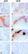
Histochemical demonstration of the presence of α4 integrin ligands in LFA-1+/+ pLNs (a–d) and mLNs (e–h). The panels show staining of identical tissue sections of HEVs in the pLNs with mAbs specific for (a) VCAM-1, (b) PNAd, (c) MAdCAM-1, and (d) an isotype-matched IgG1 control mAb and similarly, in the mLNs for (e) VCAM-1, (f) PNAd, (g) MAdCAM-1, and (h) IgG1 control mAb. Original magnification: a–h, ×150.
Figure 8
Figure 8
The effect of various mAbs on the relative entry of LFA-1−/− to LFA-1+/+ lymphocytes into bone marrow. In the absence of antibody, the relative ratio is 1.1. Coinjection of α4 mAb or VCAM-1 mAb completely inhibited the migration of LFA-1−/− cells, whereas α4β7 mAb has a partial effect and MAdCAM-1 mAb is without effect.

References

    1. Butcher EC. Leukocyte-endothelial cell recognition: three (or more) steps to specificity and diversity. Cell. 1991;67:1033–1036. - PubMed
    1. Springer TA. Traffic signals for lymphocyte recirculation and leukocyte emigration: the multistep paradigm. Cell. 1994;76:301–314. - PubMed
    1. Bargatze RF, Jutila MA, Butcher EC. Distinct roles of L-selectin and integrins α4β7 and LFA-1 in lymphocyte homing to Peyer's patch-HEV in situ: the multistep model confirmed and refined. Immunity. 1995;3:99–108. - PubMed
    1. Salmi M, Jalkanen S. How do lymphocytes know where to go: current concepts and enigmas of lymphocyte homing. Adv Immunol. 1997;64:139–218. - PubMed
    1. Butcher EC, Picker LJ. Lymphocyte homing and homeostasis. Science. 1996;272:60–66. - PubMed

Publication types